LAP3 Antibodies
Background
The LAP3 gene encodes a leucine aminopeptidase, and its expression product, as a cytoplasmic metalloproteinase, is widely present in eukaryotes. This enzyme participates in the metabolic regulation of intracellular proteins and the recycling of amino acids by catalyzing the hydrolysis of leucine residues at the N-terminal of proteins. Studies have shown that LAP3 plays a significant role in the regulation of the cell cycle, and its activity changes are closely related to the occurrence and development of tumors. This gene was first identified by cDNA cloning technology in the 1990s. The tertiary structure of the protease it encodes was analyzed by X-ray crystallography in the early 21st century, revealing a typical folded conformation of metal peptidase. As an important member of the aminopeptidase family, the structural and functional studies of LAP3 provide an important molecular basis for understanding the mechanism of intracellular protein degradation, the regulation of cell proliferation, and the mechanisms of related diseases.
Structure of LAP3
Leucine aminopeptidase encoded by the LAP3 gene is a protein with a molecular weight of approximately 56.4 kDa. This molecular weight may fluctuate slightly among different species due to minor differences in amino acid sequences.
| Species | Human | Mouse | Cattle | Zebrafish | Yeast |
| Molecular Weight (kDa) | 56.4 | 56.1 | 56.6 | 55.8 | 54.9 |
| Primary Structural Differences | Conserved sequence, highly similar to other mammals | Minor amino acid variations | Slightly different oxygen affinity | Adapted for prolonged oxygen storage | Similar to human myoglobin |
The LAP3 protein is composed of 519 amino acid residues, and its primary structure forms a typical two-domain topological configuration through specific folding. The active center of this protein contains a conserved zinc ion-binding motif (GAMENEX domain), which achieves catalytic function by coordinating metal ions. The enzymatic hydrolysis reaction is mediated by two zinc ions: one zinc ion is responsible for activating water molecules for nucleophilic attack, while the other stabilizes the carboxyl oxygen atom of the substrate. The glutamic acid residues deep in the active pocket act as generalized base catalysts, while the hydrogen bond network composed of histidine and aspartic acid precisely regulates the substrate orientation, ensuring the specific recognition of leucine residues.
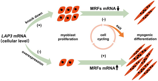 Fig. 1 Myoblast proliferation and differentiation upon LAP3 modulation.1
Key structural properties of LAP3:
- Typical α/β hydrolase folding configuration
- Dual-core zinc metal center a catalytic active site
- Conservative GAMENEX motif is responsible for the substrate specificity of recognition
- Hydrophobic pocket through hydrogen bonding network combination of precise regulation leucine residues
Functions of LAP3
The main function of the LAP3 protein is to participate in the metabolic regulation of intracellular peptide substances. In addition, it also involves a variety of cellular physiological processes, including cell cycle regulation and signal transduction.
| Function | Description |
| Peptide metabolism | Catalyze the hydrolysis of hydrophobic amino acids such as leucine at the N-terminal of proteins and participate in the final degradation step of short peptides within cells. |
| Cell cycle regulation | By hydrolyzing specific cell cycle regulatory factors, it affects the G1/S phase transition and participates in the fine regulation of the cell proliferation process. |
| Maintenance of protein homeostasis | As a supplement to the proteasome degradation pathway, it eliminates short peptides produced by normal metabolism and maintains the stability of the intracellular amino acid pool. |
| Signal transduction regulation | By modifying the activity of certain signal peptides, it indirectly affects related cellular signaling pathways, such as cellular stress responses and apoptosis processes. |
| Tumorigenesis association | The abnormal expression in a variety of cancer cells, may be affected by cell proliferation and survival pathways, involved in the occurrence and development of the tumor. |
The enzymatic kinetics curve of LAP3 exhibits typical characteristics of the Mie equation, in contrast to the biphasic curves of digestive proteases such as trypsin, which reflects its continuous response ability as an intracellular constitutive hydrolase to substrate concentration.
Applications of LAP3 and LAP3 Antibody in Literature
1. Li, Li, et al. "LAP3 contributes to IFN-γ-induced arginine depletion and malignant transformation of bovine mammary epithelial cells." BMC cancer 22.1 (2022): 864. https://doi.org/10.1186/s12885-022-09963-w
This study reveals that LAP3 mediates IFN-γ -induced arginine depletion, promoting malignant transformation of mammary epithelial cells by inhibiting ASS1 and upregulating HDAC2 and cyclin. LAP3 is highly expressed in clinical samples of breast cancer and is a potential therapeutic target.
2. Feng, Lina, et al. "Cholesterol-induced leucine aminopeptidase 3 (LAP3) upregulation inhibits cell autophagy in pathogenesis of NAFLD." Aging (Albany NY) 14.7 (2022): 3259. https://doi.org/10.18632/aging.204011
This study reveals that cholesterol can upregulate the expression of LAP3 in hepatocytes and promote the occurrence and development of non-alcoholic fatty liver disease (NAFLD) by inhibiting autophagy. Serum LAP3 levels are significantly correlated with clinical indicators such as triglycerides and glutamyl transpeptidase, and are expected to become a new biomarker for the diagnosis of NAFLD.
3. Ge, Ling, et al. "New insight into the role of the leucine aminopeptidase 3 (LAP3) in cell proliferation and myogenic differentiation in sheep embryonic myoblasts." Genes 13.8 (2022): 1438. https://doi.org/10.3390/genes13081438
This study reveals that LAP3 plays a key role in the development of myoblasts in sheep embryos. Its expression is relatively low during the proliferative phase and increases during the differentiated phase. LAP3 inhibits cell proliferation by prolonging the S phase, but promotes myotube formation and the expression of myogenic regulatory factors, thereby positively regulating terminal differentiation.
4. Worku, Destaw, and Archana Verma. "Genetic variation in bovine LAP3 and SIRT1 genes associated with fertility traits in dairy cattle." BMC Genomic Data 25.1 (2024): 32. https://doi.org/10.1186/s12863-024-01209-x
This study confirmed that SNP polymorphisms in multiple promoter regions of the LAP3 gene were significantly associated with reproductive traits such as the age at first birth and calving interval of dairy cows. Specific haplotype combinations can affect reproductive performance, indicating that LAP3 can serve as an important molecular marker for improving the reproductive capacity of dairy cows.
5. La, Yongfu, et al. "Molecular characterization and expression of SPP1, LAP3 and LCORL and their association with growth traits in sheep." Genes 10.8 (2019): 616. https://doi.org/10.3390/genes10080616
This study found that the LAP3 gene was highly expressed in the spleen, lungs, kidneys and duodenum of Hu sheep. The two mutation points (c.232C>G and c.1154C>T) were significantly correlated with growth traits such as birth weight and weight at one month of age. Among them, the c allele at the c.232C>G locus was the dominant allele and could significantly increase body weight. LAP3 can serve as an important candidate gene for improving the growth traits of sheep.
Company A: LAP3 Antibodies for Research
Company A specializes in the production of high-quality LAP3 antibodies for research and industrial applications. Our portfolio includes monoclonal antibodies tailored for ELISA, Flow Cytometry, Western blot, immunohistochemistry, and other diagnostic methodologies.
- Custom LAP3 Antibody Development: Tailor-made solutions to meet specific research requirements.
- Bulk Production: Large-scale antibody manufacturing for industry partners.
- Technical Support: Expert consultation for protocol optimization and troubleshooting.
- Aliquoting Services: Conveniently sized aliquots for long-term storage and consistent experimental outcomes.
For more details on our LAP3 antibodies, custom preparations, or technical support, contact us at email.
Reference
- Ge, Ling, et al. "New insight into the role of the leucine aminopeptidase 3 (LAP3) in cell proliferation and myogenic differentiation in sheep embryonic myoblasts." Genes 13.8 (2022): 1438. https://doi.org/10.3390/genes13081438
Anti-LAP3 antibodies
 Loading...


Hot products 
-
Mouse Anti-AFDN Recombinant Antibody (V2-58751) (CBMAB-L0408-YJ)

-
Mouse Anti-ARID3A Antibody (A4) (CBMAB-0128-YC)

-
Mouse Anti-AMACR Recombinant Antibody (CB34A) (CBMAB-CA034LY)

-
Mouse Anti-BSN Recombinant Antibody (219E1) (CBMAB-1228-CN)

-
Rat Anti-C5AR1 Recombinant Antibody (8D6) (CBMAB-C9139-LY)

-
Mouse Anti-Acetyl SMC3 (K105/K106) Recombinant Antibody (V2-634053) (CBMAB-AP052LY)

-
Mouse Anti-CD247 Recombinant Antibody (6B10.2) (CBMAB-C1583-YY)

-
Mouse Anti-CD83 Recombinant Antibody (HB15) (CBMAB-C1765-CQ)

-
Mouse Anti-BLK Recombinant Antibody (CBYY-0618) (CBMAB-0621-YY)

-
Rabbit Anti-ALK (Phosphorylated Y1278) Recombinant Antibody (D59G10) (PTM-CBMAB-0035YC)

-
Rabbit Anti-CBL Recombinant Antibody (D4E10) (CBMAB-CP0149-LY)

-
Mouse Anti-BAD (Phospho-Ser136) Recombinant Antibody (CBYY-0138) (CBMAB-0139-YY)

-
Mouse Anti-ASB9 Recombinant Antibody (1D8) (CBMAB-A0529-LY)

-
Mouse Anti-CD24 Recombinant Antibody (HIS50) (CBMAB-C10123-LY)

-
Mouse Anti-DDC Recombinant Antibody (8E8) (CBMAB-0992-YC)

-
Rabbit Anti-BAD (Phospho-Ser136) Recombinant Antibody (CAP219) (CBMAB-AP536LY)

-
Mouse Anti-ATG5 Recombinant Antibody (9H197) (CBMAB-A3945-YC)

-
Mouse Anti-ADV Recombinant Antibody (V2-503423) (CBMAB-V208-1364-FY)

-
Mouse Anti-AGK Recombinant Antibody (V2-258056) (CBMAB-M0989-FY)

-
Mouse Anti-4-Hydroxynonenal Recombinant Antibody (V2-502280) (CBMAB-C1055-CN)

- AActivation
- AGAgonist
- APApoptosis
- BBlocking
- BABioassay
- BIBioimaging
- CImmunohistochemistry-Frozen Sections
- CIChromatin Immunoprecipitation
- CTCytotoxicity
- CSCostimulation
- DDepletion
- DBDot Blot
- EELISA
- ECELISA(Cap)
- EDELISA(Det)
- ESELISpot
- EMElectron Microscopy
- FFlow Cytometry
- FNFunction Assay
- GSGel Supershift
- IInhibition
- IAEnzyme Immunoassay
- ICImmunocytochemistry
- IDImmunodiffusion
- IEImmunoelectrophoresis
- IFImmunofluorescence
- IGImmunochromatography
- IHImmunohistochemistry
- IMImmunomicroscopy
- IOImmunoassay
- IPImmunoprecipitation
- ISIntracellular Staining for Flow Cytometry
- LALuminex Assay
- LFLateral Flow Immunoassay
- MMicroarray
- MCMass Cytometry/CyTOF
- MDMeDIP
- MSElectrophoretic Mobility Shift Assay
- NNeutralization
- PImmunohistologyp-Paraffin Sections
- PAPeptide Array
- PEPeptide ELISA
- PLProximity Ligation Assay
- RRadioimmunoassay
- SStimulation
- SESandwich ELISA
- SHIn situ hybridization
- TCTissue Culture
- WBWestern Blot
