MICB Antibodies
Background
The MICB gene encodes a protein belonging to MHC Class I related molecules and is mainly expressed on the surface of immune cells. This protein, as a ligand, can bind to the NKG2D receptor on the surface of natural killer cells and T cells, thereby activating cytotoxic immune responses and eliminating abnormal cells. During the process of antiviral immunity and tumor immunity surveillance, MICB marks infected or malignant transformed cells by upregulating their expression, guiding the immune system to carry out precise attacks. This gene was discovered through the Human Genome Project in 1994. Its unique non-classical MHC molecular structure provides an important model for the study of immune recognition mechanisms. The related research results not only deepen the understanding of the connection mechanism between innate immunity and adaptive immunity, but also lay a molecular foundation for the development of new immunotherapies.
Structure of MICB
MICB is an immunomodulatory protein with a molecular weight of approximately 42 kDa, and this value varies slightly among different species.
| Species | Human | Mouse | Rhesus monkey | Bovine |
| Molecular Weight (kDa) | 42.0 | 41.8 | 41.9 | 42.1 |
| Primary Structural Differences | Typical MHC class I-like structures | There is sequence variation in the α3 domain | Highly homologous to humans | Extracellular region conservative is higher |
This protein is encoded by MHC Class I chain-related gene B and has a typical MHC-like folding structure, containing three extracellular domains: α1, α2, and α3. The α1 and α2 domains of MICB jointly form a conserved antigen-binding slot. However, unlike classical MHC molecules, this domain does not bind to the peptide but directly interacts with the NKG2D receptor. Its α3 domain is similar to the folding of immunoglobulins and is anchored to the cell membrane through the transmembrane region. The intracellular region of proteins contains key signal transduction modalities that can mediate the upregulation of cell surface expression under stress conditions, thereby initiating an immune response.
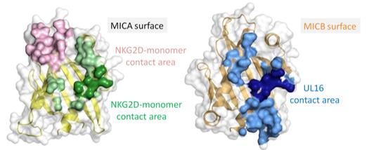 Fig. 1 Comparative Structures of the MICA and MICB α1α2 Platform Domains.1
Key structural properties of MICB:
- Typical MHC class I-like folding configuration
- It consists of three extracellular domains, α1, α2 and α3
- The α1-α2 domain forms an interface that specifically binds to the NKG2D receptor
- Transmembrane regions anchor proteins to the surface of immune cells
Functions of MICB
The main function of the MICB gene is to participate in the activation and regulation of immune responses, and it also plays a role in various immune-related physiological processes.
| Function | Description |
| Immune activation | As a ligand of the NKG2D receptor, it activates the immune response of natural killer cells and cytotoxic T cells. |
| Stress signal indication | Malignant transformation occur in infected cells or raised expression, for the immune system to identify clear mark abnormal cells. |
| Anti-tumor immunity | By inducing immune cells to kill tumor cells, it participates in the process of tumor immune surveillance. |
| Antiviral infection | Present on the surface of virus-infected cells, instructing immune cells to specifically clear infected cells. |
| Regulation of immune homeostasis | Participate in maintaining the balance between immune tolerance and immune activation in normal tissues. |
The binding of MICB to NKG2D exhibits high affinity characteristics. Compared with the specific recognition of classical MHC-peptide complexes, its mechanism of action is more direct and efficient, which enables it to rapidly initiate anti-pathogen and anti-tumor defenses in the early stage of immune response.
Applications of MICB and MICB Antibody in Literature
1. Feng, Qingyang, et al. "High MICB expression as a biomarker for good prognosis of colorectal cancer." Journal of Cancer Research and Clinical Oncology 146.6 (2020): 1405-1413. https://doi.org/10.1007/s00432-020-03159-0
This study confirmed through two independent cocohort studies that high expression of MICB is an independent prognostic protective factor for colorectal cancer, significantly associated with better overall survival of patients, and is conducive to more precise prognostic risk stratification.
2. Faridah, Imaniar Noor, et al. "Genetic association studies of MICB and PLCE1 with severity of dengue in Indonesian and Taiwanese populations." Diagnostics 13.21 (2023): 3365. https://doi.org/10.3390/diagnostics13213365
Research has found that the variant rs3132468 of the MHC gene MICB is significantly associated with dengue shock syndrome, while the variant of the PLCE1 gene is related to severe secondary infection. This provides a basis for genetic screening of severe dengue fever.
3. Baek, In-Cheol, et al. "MICB allele genotyping on microarrays by improving the specificity of extension primers." Plos one 10.11 (2015): e0142467. https://doi.org/10.1371/journal.pone.0142467
This study successfully established a MICB genotyping method based on microarray allele-specific primer extension (ASPE), and the typing results were completely consistent with the sequencing method, which is suitable for large-scale population genetics research.
4. Whitehorn, James, et al. "Genetic variants of MICB and PLCE1 and associations with non-severe dengue." PloS one 8.3 (2013): e59067. https://doi.org/10.1371/journal.pone.0059067
Research has confirmed that specific variations in the MICB and PLCE1 genes (rs3132468 and rs3740360) not only increase the risk of dengue shock syndrome but are also significantly associated with common dengue fever and infantichild infections, deepening the understanding of the pathogenic mechanism of dengue fever.
5. Whitehorn, James, et al. "Genetic variants of MICB and PLCE1 and associations with the laboratory features of dengue." BMC infectious diseases 17.1 (2017): 412. https://doi.org/10.1186/s12879-017-2514-6
Research has found that although MICB and PLCE1 gene variations are known to be associated with the clinical severity of dengue fever, their specific mechanisms of action remain unclear. Analysis shows that these variations have no significant association with key laboratory indicators such as viral load and platelet count.
Creative Biolabs: MICB Antibodies for Research
Creative Biolabs specializes in the production of high-quality MICB antibodies for research and industrial applications. Our portfolio includes monoclonal antibodies tailored for ELISA, Flow Cytometry, Western blot, immunohistochemistry, and other diagnostic methodologies.
- Custom MICB Antibody Development: Tailor-made solutions to meet specific research requirements.
- Bulk Production: Large-scale antibody manufacturing for industry partners.
- Technical Support: Expert consultation for protocol optimization and troubleshooting.
- Aliquoting Services: Conveniently sized aliquots for long-term storage and consistent experimental outcomes.
For more details on our MICB antibodies, custom preparations, or technical support, contact us at email.
Reference
- Müller, Steffen, et al. "Structure of the HCMV UL16-MICB complex elucidates select binding of a viral immunoevasin to diverse NKG2D ligands." PLoS pathogens 6.1 (2010): e1000723. https://doi.org/10.1371/journal.ppat.1000723
Anti-MICB antibodies
 Loading...


Hot products 
-
Rat Anti-ADAM10 Recombinant Antibody (V2-179741) (CBMAB-A1103-YC)

-
Mouse Anti-CD33 Recombinant Antibody (P67.6) (CBMAB-C10189-LY)

-
Mouse Anti-CCNH Recombinant Antibody (CBFYC-1054) (CBMAB-C1111-FY)

-
Mouse Anti-FN1 Monoclonal Antibody (71) (CBMAB-1241CQ)

-
Rat Anti-CD63 Recombinant Antibody (7G4.2E8) (CBMAB-C8725-LY)

-
Mouse Anti-GIPC2 Recombinant Antibody (10) (CBMAB-G0476-LY)

-
Rat Anti-EPO Recombinant Antibody (16) (CBMAB-E1578-FY)

-
Rabbit Anti-AKT2 (Phosphorylated S474) Recombinant Antibody (V2-556130) (PTM-CBMAB-0605LY)

-
Mouse Anti-APP Recombinant Antibody (DE2B4) (CBMAB-1122-CN)

-
Mouse Anti-ATP1B3 Recombinant Antibody (1E9) (CBMAB-A4021-YC)

-
Mouse Anti-BRCA2 Recombinant Antibody (CBYY-1728) (CBMAB-2077-YY)

-
Mouse Anti-AFDN Recombinant Antibody (V2-58751) (CBMAB-L0408-YJ)

-
Mouse Anti-ADAM29 Recombinant Antibody (V2-179787) (CBMAB-A1149-YC)

-
Mouse Anti-ARHGAP5 Recombinant Antibody (54/P190-B) (CBMAB-P0070-YC)

-
Rabbit Anti-ADRA1A Recombinant Antibody (V2-12532) (CBMAB-1022-CN)

-
Mouse Anti-DLL4 Recombinant Antibody (D1090) (CBMAB-D1090-YC)

-
Mouse Anti-ENO2 Recombinant Antibody (85F11) (CBMAB-0276CQ)

-
Mouse Anti-AZGP1 Recombinant Antibody (CBWJZ-007) (CBMAB-Z0012-WJ)

-
Mouse Anti-ATG5 Recombinant Antibody (9H197) (CBMAB-A3945-YC)

-
Mouse Anti-CORO1A Recombinant Antibody (4G10) (V2LY-1206-LY806)

- AActivation
- AGAgonist
- APApoptosis
- BBlocking
- BABioassay
- BIBioimaging
- CImmunohistochemistry-Frozen Sections
- CIChromatin Immunoprecipitation
- CTCytotoxicity
- CSCostimulation
- DDepletion
- DBDot Blot
- EELISA
- ECELISA(Cap)
- EDELISA(Det)
- ESELISpot
- EMElectron Microscopy
- FFlow Cytometry
- FNFunction Assay
- GSGel Supershift
- IInhibition
- IAEnzyme Immunoassay
- ICImmunocytochemistry
- IDImmunodiffusion
- IEImmunoelectrophoresis
- IFImmunofluorescence
- IGImmunochromatography
- IHImmunohistochemistry
- IMImmunomicroscopy
- IOImmunoassay
- IPImmunoprecipitation
- ISIntracellular Staining for Flow Cytometry
- LALuminex Assay
- LFLateral Flow Immunoassay
- MMicroarray
- MCMass Cytometry/CyTOF
- MDMeDIP
- MSElectrophoretic Mobility Shift Assay
- NNeutralization
- PImmunohistologyp-Paraffin Sections
- PAPeptide Array
- PEPeptide ELISA
- PLProximity Ligation Assay
- RRadioimmunoassay
- SStimulation
- SESandwich ELISA
- SHIn situ hybridization
- TCTissue Culture
- WBWestern Blot
