MSLN Antibodies
Background
The MSLN gene encodes a glycoprotein known as mesothelin. This protein is typically anchored to the surface membrane of normal mesothelial cells and is mainly distributed in the pleura, peritoneum, and pericardium. Mesothelin is involved in cell adhesion and signal transduction, and may affect cell proliferation and survival in certain physiological processes. This gene is highly expressed in various malignant tumors, especially in mesothelioma, pancreatic cancer, and ovarian cancer. Therefore, it has become an important biomarker for tumor-targeted therapy and diagnostic research. Since its first identification in 1992, new treatment strategies such as antibody-drug conjugates targeting mesothelin and CAR-T cell therapy have entered the clinical development stage. The research on its structure and function is continuously advancing the progress of cancer precision medicine.
Structure of MSLN
The mesothelin protein encoded by the MSLN gene is a glycosylphosphatidylinositol-anchored glycoprotein with a molecular weight of approximately 40 kDa. Its molecular weight may vary depending on different splicing variants or glycosylation modifications. The core domain of this protein is highly conserved, but there are subtle differences in amino acid sequences among different species.
| Species | Human | Mouse | Rhesus monkey |
| Molecular Weight (kDa) | ~40 | ~38 | ~40 |
| Primary Structural Differences | Containing the signal peptide, the N-terminal of MSLN and the GPI anchoring sequence | High sequence homology and conserved functional domains | Highly homologous to human MSLN, it is a commonly used preclinical model |
The primary structure of desmin protein consists of approximately 622 amino acid residues, and it folds to form multiple domains. The N-terminal domain of the protein (MSLN-N, approximately 30 kDa) plays a role in cell adhesion and interacts with ligands such as CA125 through its tertiary structure. The C-terminal is anchored to the cell membrane via GPI. The secondary structure is mainly composed of β-sheet and α-helix, which together maintain its stable spatial conformation and enable its biological functions.
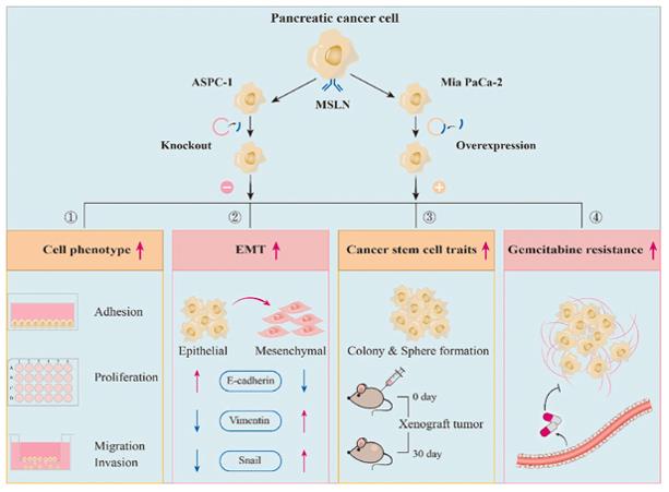 Fig. 1 MSLN Drives Aggression, Stemness, and Chemoresistance in Pancreatic Cancer.1
Key structural properties of MSLN:
- Transmembrane glycoprotein structure with GPI anchoring
- The N-terminal domain (MSLN-N) mediates cell adhesion and recognition
- The C-terminal glycosyl phosphatidylinositol anchoring domain is responsible for membrane localization
- Rich in glycosylation modification sites on the surface
Functions of MSLN
The MSLN gene encodes the protein (mesothelin), which mainly functions in normal physiological conditions to participate in cell adhesion and signal transduction. Its dysfunction is closely related to various pathological processes, and it plays a crucial role in tumor occurrence and development.
| Function | Description |
| Cell Adhesion | By interacting with molecules such as CA125/MUC16 through its N-terminal domain, it mediates the recognition and adhesion between cells. |
| Cell Proliferation and Survival | Regulates the signaling pathways involved in cell growth, proliferation and apoptosis, and affects tissue homeostasis. |
| Tumor Progression and Metastasis | Overexpressed in various cancers, it enhances the adhesion of cancer cells to the stroma, promoting tumor invasion, metastasis and peritoneal dissemination. |
| Chemotherapy Resistance | This refers to the resistance of tumor cells to specific chemotherapy drugs, and it is one of the factors that affect the treatment outcome. |
| Immune Regulation | Its expression level can affect the tumor microenvironment and can serve as a specific target for immunotherapy (such as CAR-T, antibody drugs). |
Unlike typical secretory proteins, desmosomal proteins are anchored to the cell membrane through GPI. Their functions are highly "position-specific", and they mainly exert their functions by interacting with ligands on the adjacent cell surface, which makes them particularly important in the local invasion and metastasis of tumors.
Applications of MSLN and MSLN Antibody in Literature
1. Hu, Jili, et al. "MSLN induced EMT, cancer stem cell traits and chemotherapy resistance of pancreatic cancer cells." Heliyon 10.8 (2024). https://doi.org/10.1016/j.heliyon.2024.e29210
Studies have shown that the high expression of MSLN in pancreatic cancer cells leads to chemotherapy resistance by enhancing migration, invasion, epithelial-mesenchymal transition and stem cell properties. Targeting MSLN may reverse these processes and become a potential therapeutic strategy.
2. Schoutrop, Esther, et al. "Tuned activation of MSLN-CAR T cells induces superior antitumor responses in ovarian cancer models." Journal for immunotherapy of cancer 11.2 (2023): e005691. https://doi.org/10.1136/jitc-2022-005691
The research found that in ovarian cancer models, the MSLN-CAR-T (M1xx) with a single ITAM mutation in the CD3ζ chain demonstrated significantly enhanced anti-tumor activity, durability, and reduced T-cell exhaustion compared to the traditional second-generation structure, providing a new strategy for the treatment of solid tumors.
3. Li, Yike, et al. "MSLN correlates with immune infiltration and chemoresistance as a prognostic biomarker in ovarian cancer." Frontiers in oncology 12 (2022): 830570. https://doi.org/10.3389/fonc.2022.830570
The study found that MSLN is highly expressed in ovarian cancer and is associated with poor prognosis. It may cause the deterioration of ovarian cancer prognosis by inhibiting immune activation and promoting multiple pathways of chemotherapy resistance, suggesting that it is a potential therapeutic target.
4. Schoutrop, Esther, et al. "Trogocytosis and fratricide killing impede MSLN-directed CAR T cell functionality." Oncoimmunology 11.1 (2022): 2093426. https://doi.org/10.1080/2162402X.2022.2093426
The research has found that MSLN-CAR-T cells with CD28 co-stimulation (M28z) have a stronger oncolytic ability compared to those with 4-1BB co-stimulation (MBBz). The bottleneck lies in the self-cannibalization caused by CAR-mediated endocytosis and the antigen heterogeneity, and the upregulation of LAG-3 will weaken the function of T cells.
5. Zeng, Wuyi, et al. "A novel PD-L1-Containing MSLN targeting vaccine for lung cancer immunotherapy." Frontiers in Immunology 13 (2022): 925217. https://doi.org/10.3389/fimmu.2022.925217
This study developed a novel MSLN-PDL1-GMCSF vaccine, which can effectively induce mice to produce strong anti-PD-L1 antibodies and specific cytotoxic T cell responses, and significantly inhibit the growth of lung cancer. When combined with PD-1 blockade, it can further enhance the synergistic anti-tumor efficacy.
Creative Biolabs: MSLN Antibodies for Research
Creative Biolabs specializes in the production of high-quality MSLN antibodies for research and industrial applications. Our portfolio includes monoclonal antibodies tailored for ELISA, Flow Cytometry, Western blot, immunohistochemistry, and other diagnostic methodologies.
- Custom MSLN Antibody Development: Tailor-made solutions to meet specific research requirements.
- Bulk Production: Large-scale antibody manufacturing for industry partners.
- Technical Support: Expert consultation for protocol optimization and troubleshooting.
- Aliquoting Services: Conveniently sized aliquots for long-term storage and consistent experimental outcomes.
For more details on our MSLN antibodies, custom preparations, or technical support, contact us at email.
Reference
- Hu, Jili, et al. "MSLN induced EMT, cancer stem cell traits and chemotherapy resistance of pancreatic cancer cells." Heliyon 10.8 (2024). Distributed under Open Access license CC BY 4.0, without modification. https://doi.org/10.1016/j.heliyon.2024.e29210
Anti-MSLN antibodies
 Loading...


Hot products 
-
Mouse Anti-ATP5F1A Recombinant Antibody (51) (CBMAB-A4043-YC)

-
Mouse Anti-ATG5 Recombinant Antibody (9H197) (CBMAB-A3945-YC)

-
Mouse Anti-AMOT Recombinant Antibody (CBYC-A564) (CBMAB-A2552-YC)

-
Mouse Anti-FN1 Monoclonal Antibody (D6) (CBMAB-1240CQ)

-
Mouse Anti-AKR1B1 Antibody (V2-2449) (CBMAB-1001CQ)

-
Mouse Anti-FLI1 Recombinant Antibody (CBXF-0733) (CBMAB-F0435-CQ)

-
Mouse Anti-ARIH1 Recombinant Antibody (C-7) (CBMAB-A3563-YC)

-
Mouse Anti-FN1 Monoclonal Antibody (71) (CBMAB-1241CQ)

-
Mouse Anti-APCS Recombinant Antibody (CBYC-A663) (CBMAB-A3054-YC)

-
Mouse Anti-CCT6A/B Recombinant Antibody (CBXC-0168) (CBMAB-C5570-CQ)

-
Mouse Anti-CDKL5 Recombinant Antibody (CBFYC-1629) (CBMAB-C1689-FY)

-
Mouse Anti-FeLV g27 Recombinant Antibody (1) (CBMAB-V208-1714-FY)

-
Mouse Anti-ATP1B1 Recombinant Antibody (E4) (CBMAB-0463-LY)

-
Rabbit Anti-ALK (Phosphorylated Y1278) Recombinant Antibody (D59G10) (PTM-CBMAB-0035YC)

-
Mouse Anti-CCDC6 Recombinant Antibody (CBXC-0106) (CBMAB-C5397-CQ)

-
Rat Anti-CD34 Recombinant Antibody (MEC 14.7) (CBMAB-C10196-LY)

-
Mouse Anti-GLP1R Recombinant Antibody (4F3) (CBMAB-G0521-LY)

-
Mouse Anti-CTCF Recombinant Antibody (CBFYC-2371) (CBMAB-C2443-FY)

-
Mouse Anti-FOXA3 Recombinant Antibody (2A9) (CBMAB-0377-YC)

-
Mouse Anti-ENO1 Recombinant Antibody (8G8) (CBMAB-E1329-FY)

- AActivation
- AGAgonist
- APApoptosis
- BBlocking
- BABioassay
- BIBioimaging
- CImmunohistochemistry-Frozen Sections
- CIChromatin Immunoprecipitation
- CTCytotoxicity
- CSCostimulation
- DDepletion
- DBDot Blot
- EELISA
- ECELISA(Cap)
- EDELISA(Det)
- ESELISpot
- EMElectron Microscopy
- FFlow Cytometry
- FNFunction Assay
- GSGel Supershift
- IInhibition
- IAEnzyme Immunoassay
- ICImmunocytochemistry
- IDImmunodiffusion
- IEImmunoelectrophoresis
- IFImmunofluorescence
- IGImmunochromatography
- IHImmunohistochemistry
- IMImmunomicroscopy
- IOImmunoassay
- IPImmunoprecipitation
- ISIntracellular Staining for Flow Cytometry
- LALuminex Assay
- LFLateral Flow Immunoassay
- MMicroarray
- MCMass Cytometry/CyTOF
- MDMeDIP
- MSElectrophoretic Mobility Shift Assay
- NNeutralization
- PImmunohistologyp-Paraffin Sections
- PAPeptide Array
- PEPeptide ELISA
- PLProximity Ligation Assay
- RRadioimmunoassay
- SStimulation
- SESandwich ELISA
- SHIn situ hybridization
- TCTissue Culture
- WBWestern Blot
