NFIA Antibodies
Background
The NFIA gene encodes nuclear factor IA-type transcription factor, which, as A key molecule regulating glial cell differentiation, is widely involved in the development process of the central nervous system. This protein regulates the expression of downstream target genes by binding to specific DNA sequences. It not only affects the fate determination of glial cells but also participates in the formation and maintenance of the blood-brain barrier. Research has found that mutations in the NFIA gene are closely related to central nervous system malformations (such as corpus callosum hypoplasia) and cognitive dysfunction. The research on its molecular mechanism provides an important perspective for revealing the pathological mechanism of neurodevelopmental disorders, especially having key scientific value in the field of glial cell-neuron interaction.
Structure of NFIA
The molecular weight of NFIA protein is approximately 55-65 kDa, and its precise value varies among different transcript isomers and species.
| Species | Human | Mouse | Rat | Zebrafish |
| Molecular Weight (kDa) | 62 | 58 | 59 | 54 |
| Primary Structural Differences | Contain highly conserved DNA binding domain and transcription regulation area | Homologous DNA binding domain is extremely high | Species-specific variations exist in the regulatory regions | Typical structure have the vertebrate NFIA family |
This protein is composed of approximately 500 amino acids, and its primary structure forms a typical helical-ring-helical DNA-binding motif. The tertiary structure of proteins forms functional homologous/heterodimers through dimerization domains, thereby specifically recognizing the TGGCANNTGCC sequence. The structure contains nuclear localization signals to ensure its location in the nucleus, while the C-terminal transcriptional activation domain regulates the chromatin state of the target gene by recruiting histone modification complexes.
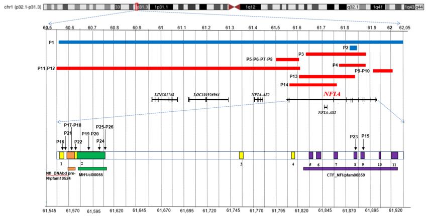 Fig. 1 Overview of NFIA pathogenic variants.1
Key structural properties of NFIA:
- Contains highly conserved DNA binding domains
- Form a specific dimerization interface
- Equipped with nuclear localization signal and transcription regulation module
Functions of NFIA
The core function of the NFIA gene is to regulate the development of the nervous system and cell differentiation. In addition, this gene is also involved in multiple physiological and pathological processes such as organ formation and tumorigenesis.
| Function | Description |
| Neurodevelopmental regulation | By activating specific target genes, it guides neural precursor cells to differentiate into astrocyte lineages, promoting the normal development of the cerebral cortex. |
| Cell differentiation determination | With various transcription factors synergy, glial cell fate decisions, and participate in the epithelial - interstitial transformation process. |
| Organ morphogenesis | In the heart, kidneys organs such as the development regulation of related gene expression, mature influence organization structure and function. |
| Tumorigenesis influence | The expression is abnormal in some gliomas and epithelial tumors, which may play a role by changing the cell proliferation signaling pathway. |
| Chromatin remodeling | By recruiting histone modification complexes to alter the chromatin open state, a tissue-specific gene expression pattern is established. |
The heterodimers formed by NFIA and its family members can recognize specific DNA sequences. Their regulatory patterns show environmental dependence, capable of both activating and inhibiting target genes, demonstrating their fine regulatory characteristics during development.
Applications of NFIA and NFIA Antibody in Literature
1. Bertini, Veronica, et al. "Phenotypic spectrum of NFIA haploinsufficiency: two additional cases and review of the literature." Genes 13.12 (2022): 2249. https://doi.org/10.3390/genes13122249
Research has revealed that haploid insufficiency of the NFIA gene can lead to the core phenotype of 1p32p31 deletion syndrome, with typical features including macrocephaly, special facial signs, abnormal thumbs, and often accompanied by intellectual development disorders or neurodevelopmental abnormalities. MRI and ultrasound examinations can assist in diagnosis, but genetic testing is ultimately required for confirmation.
2. Dini, Gianluca, et al. "NFIA haploinsufficiency: case series and literature review." Frontiers in Pediatrics 11 (2023): 1292654. https://doi.org/10.3389/fped.2023.1292654
Research reveals that NFIA haploidy deficiency is a rare neurodevelopmental disorder, with typical features including intellectual disability, macrocephaly and craniofacial abnormalities. Renal ultrasound and MRI can provide clues, and ultimately, genetic testing is needed for a confirmed diagnosis.
3. Kong, Shuo, et al. "Cell-specific NFIA upregulation promotes epileptogenesis by TRPV4-mediated astrocyte reactivity." Journal of Neuroinflammation 20.1 (2023): 247. https://doi.org/10.1186/s12974-023-02909-4
Research has found that in epilepsy models, hippocampal astrocytes specifically and highly express NFIA, which in turn upregulates TRPV4 channels, intensifies inflammatory responses and promotes epileptic seizures. Inhibiting NFIA can weaken this process, providing a new target for treatment.
4. Keeley, Patrick W., et al. "NFIA is critical for AII amacrine cell production: selective bipolar cell dependencies and diminished ERG." Journal of Neuroscience 43.49 (2023): 8367-8384. https://doi.org/10.1523/JNEUROSCI.1099-23.2023
Studies have shown that the NFIA gene is the key to the differentiation and survival of AII cells without long processes in the mouse retina. Knockout of this gene will lead to the specific deletion of such cells and cause a reduction in secondary bipolar cells as well as abnormal electrophysiological functions of the retina.
5. Uehara, Tomoko, et al. "Recurrent NFIA K125E substitution represents a loss-of-function allele: Sensitive in vitro and in vivo assays for nontruncating alleles." American journal of medical genetics Part A 185.7 (2021): 2084-2093. https://doi.org/10.1002/ajmg.a.62226
Research has found that a novel missense mutation (K125E) in the NFIA gene can lead to typical neurodevelopmental disorders. It was confirmed through fruit fly, zebrafish models and cell experiments that this mutation caused the loss of function of the NFIA protein, affecting the development of brain axons and gene regulation.
Creative Biolabs: NFIA Antibodies for Research
Creative Biolabs specializes in the production of high-quality NFIA antibodies for research and industrial applications. Our portfolio includes monoclonal antibodies tailored for ELISA, Flow Cytometry, Western blot, immunohistochemistry, and other diagnostic methodologies.
- Custom NFIA Antibody Development: Tailor-made solutions to meet specific research requirements.
- Bulk Production: Large-scale antibody manufacturing for industry partners.
- Technical Support: Expert consultation for protocol optimization and troubleshooting.
- Aliquoting Services: Conveniently sized aliquots for long-term storage and consistent experimental outcomes.
For more details on our NFIA antibodies, custom preparations, or technical support, contact us at email.
Reference
- Bertini, Veronica, et al. "Phenotypic spectrum of NFIA haploinsufficiency: two additional cases and review of the literature." Genes 13.12 (2022): 2249. https://doi.org/10.3390/genes13122249
Anti-NFIA antibodies
 Loading...


Hot products 
-
Mouse Anti-ACE2 Recombinant Antibody (V2-179293) (CBMAB-A0566-YC)

-
Rabbit Anti-CCN1 Recombinant Antibody (CBWJC-3580) (CBMAB-C4816WJ)

-
Mouse Anti-ELAVL4 Recombinant Antibody (6B9) (CBMAB-1132-YC)

-
Mouse Anti-FPR2 Recombinant Antibody (1D6) (CBMAB-F2628-CQ)

-
Mouse Anti-GLP1R Recombinant Antibody (4F3) (CBMAB-G0521-LY)

-
Mouse Anti-CD59 Recombinant Antibody (CBXC-2097) (CBMAB-C4421-CQ)

-
Rat Anti-CCR2 Recombinant Antibody (475301) (CBMAB-C1338-LY)

-
Mouse Anti-ADGRE5 Recombinant Antibody (V2-360335) (CBMAB-C2088-CQ)

-
Mouse Anti-H1-0 Recombinant Antibody (CBFYH-0565) (CBMAB-H1451-FY)

-
Human Anti-SARS-CoV-2 Spike Recombinant Antibody (CR3022) (CBMAB-CR014LY)

-
Rabbit Anti-DLK1 Recombinant Antibody (9D8) (CBMAB-D1061-YC)

-
Mouse Anti-AAV-5 Recombinant Antibody (V2-503417) (CBMAB-V208-1369-FY)

-
Mouse Anti-CDK7 Recombinant Antibody (CBYY-C1783) (CBMAB-C3221-YY)

-
Mouse Anti-ASB9 Recombinant Antibody (1D8) (CBMAB-A0529-LY)

-
Rabbit Anti-ALOX5AP Recombinant Antibody (CBXF-1219) (CBMAB-F0750-CQ)

-
Mouse Anti-CECR2 Recombinant Antibody (CBWJC-2465) (CBMAB-C3533WJ)

-
Mouse Anti-ACTB Recombinant Antibody (V2-179553) (CBMAB-A0870-YC)

-
Mouse Anti-BMI1 Recombinant Antibody (CBYC-P026) (CBMAB-P0108-YC)

-
Mouse Anti-CHRNA9 Recombinant Antibody (8E4) (CBMAB-C9161-LY)

-
Mouse Anti-CTCF Recombinant Antibody (CBFYC-2371) (CBMAB-C2443-FY)

- AActivation
- AGAgonist
- APApoptosis
- BBlocking
- BABioassay
- BIBioimaging
- CImmunohistochemistry-Frozen Sections
- CIChromatin Immunoprecipitation
- CTCytotoxicity
- CSCostimulation
- DDepletion
- DBDot Blot
- EELISA
- ECELISA(Cap)
- EDELISA(Det)
- ESELISpot
- EMElectron Microscopy
- FFlow Cytometry
- FNFunction Assay
- GSGel Supershift
- IInhibition
- IAEnzyme Immunoassay
- ICImmunocytochemistry
- IDImmunodiffusion
- IEImmunoelectrophoresis
- IFImmunofluorescence
- IGImmunochromatography
- IHImmunohistochemistry
- IMImmunomicroscopy
- IOImmunoassay
- IPImmunoprecipitation
- ISIntracellular Staining for Flow Cytometry
- LALuminex Assay
- LFLateral Flow Immunoassay
- MMicroarray
- MCMass Cytometry/CyTOF
- MDMeDIP
- MSElectrophoretic Mobility Shift Assay
- NNeutralization
- PImmunohistologyp-Paraffin Sections
- PAPeptide Array
- PEPeptide ELISA
- PLProximity Ligation Assay
- RRadioimmunoassay
- SStimulation
- SESandwich ELISA
- SHIn situ hybridization
- TCTissue Culture
- WBWestern Blot
