ORAI1 Antibodies
Background
ORAI1 is a calcium ion channel protein located in the cytoplasmic membrane, mainly present in various vertebrate cells such as immune cells and muscle cells. As the core subunit of calcium release-activated calcium channels, this protein is activated after the depletion of the intracellular calcium pool and mediates calcium ion influx, thereby regulating key physiological processes such as gene expression, cell proliferation, and immune responses. T-cell activation is highly dependent on the calcium signal mediated by ORAI1, and its dysfunction is closely related to immune deficiency diseases. In 2006, multiple research teams first identified ORAI1 as the key molecule of calcium release-activated calcium channels, which laid an important foundation for understanding the mechanism of calcium signal regulation. The interaction between ORAI1 and other proteins has become an important model for studying calcium signal transduction, ion channel regulation, and related disease mechanisms, greatly promoting our understanding of the cell signal network and precise treatment strategies.
Structure of ORAI1
ORAI1 is a multi-spanning membrane protein with a monomer molecular weight of approximately 33 kDa. The molecular weight of ORAI1 varies among different species, mainly due to the changes in the length and composition of the amino acid sequence.
| Species | Human | Mouse | Rat | Zebrafish | Fruit fly |
| Molecular Weight (kDa) | 33.0 | 32.8 | 32.9 | 33.5 | 30.2 |
| Primary Structural Differences | Classical calcium release-activated calcium channel subunit | Highly homologous to humans, with conserved function | Similar sequences, consistent calcium gating characteristics | Presence of fish-specific sequence insertions | Simpler structure, an important model for studying calcium signaling |
The ORAI1 protein consists of 301 amino acids and its three-dimensional structure is in the form of a hexamer assembly. It forms a central pore through four transmembrane α helices. Each subunit of this protein contains a characteristic transmembrane domain, among which the extracellular loop between the first and second transmembrane helices has a cluster of negatively charged amino acids, which plays a key role in the selectivity for calcium ions. The glutamic acid residue within the first transmembrane helix constitutes an ion-selective filter, determining the high specificity for calcium ions. The N-terminal and C-terminal of the ORAI1 protein are both located on the cytoplasmic side. The N-terminal contains a key domain for binding with the STIM1 protein, while the C-terminal participates in the gating regulation of the channel. The second and third transmembrane helix connection loop region also participates in the channel activation process mediated by STIM1. This unique structural design enables ORAI1 to sense the depletion signal of the endoplasmic reticulum calcium pool and achieve precise calcium ion gating.
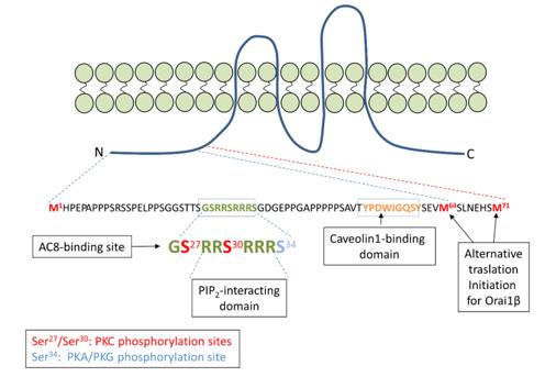 Fig. 1 Amino acid sequence of the N-terminal region unique to Orai1α.1
Key structural properties of ORAI1:
- A hexameric channel formed by four transmembrane α helices
- A central pore with a cluster of negatively charged amino acids
- Glutamate in the first transmembrane helix acts as an ion-selective filter
- The intracellular N-terminal and C-terminal domains are involved in the gating regulation mediated by STIM1
Functions of ORAI1
ORAI1, as the core subunit that activates calcium channels through calcium release, primarily functions to mediate the influx of calcium ions after the depletion of the intracellular calcium pool, maintaining the stability of cytoplasmic calcium signals.
| Function | Description |
| Calcium ion influx | After the endoplasmic reticulum calcium depletion, it binds to STIM1 protein and is activated, mediating the entry of extracellular calcium ions into the cytoplasm. |
| Immune synapse formation | Through local calcium signals, it regulates the downstream signals of the T cell receptor, promoting the maturation and stability of the immune synapse. |
| Gene Transcription Regulation | Calcium influx activates the calcineurin-NFAT pathway, driving the expression of cytokines and other related genes. |
| Cell Proliferation and Differentiation | Maintaining calcium oscillation signals and regulating the clonal expansion and functional differentiation of various immune cells such as T cells and mast cells. |
| Muscle Function Support | It mediates calcium ion influx in skeletal muscles and cardiac muscles, participating in the excitation-contraction coupling process and the development of muscles. |
Unlike the classical oxygen-binding proteins, the calcium current-voltage relationship of ORAI1 exhibits an inward rectification feature. Under negative potentials, the influx of calcium ions is significantly enhanced, while under positive potentials, the current weakens. This indicates its high selectivity for calcium ions and its unique gating characteristics during the depolarization process of the cell.
Applications of ORAI1 and ORAI1 Antibody in Literature
1. Sánchez-Collado, José, José J. López, and Juan A. Rosado. "The Orai1-AC8 interplay: how breast cancer cells escape from orai1 channel inactivation." Cells 10.6 (2021): 1308. https://doi.org/10.3390/cells10061308
The article indicates that adenylate cyclase 8 (AC8) interacts with the calcium channel Orai1α, regulating calcium signals and the cAMP/PKA pathway. In breast cancer cells, overexpression of AC8 disrupts this regulation, enhances calcium influx, and promotes cancer cell proliferation and migration, revealing a new mechanism for tumor occurrence.
2. Beech, David J. "Orai1 calcium channels in the vasculature." Pflügers Archiv-European Journal of Physiology 463.5 (2012): 635-647. https://doi.org/10.1007/s00424-012-1090-2
The research has found that the T-cell calcium channel Orai1 is also expressed in vascular smooth muscle and endothelial cells, and participates in calcium signal regulation. Orai1 plays a crucial role in vascular remodeling and contraction, and may be a new target for vascular function regulation.
3. Tao, Yu, et al. "Enhanced Orai1-mediated store-operated Ca2+ channel/calpain signaling contributes to high glucose-induced podocyte injury." Journal of Biological Chemistry 298.6 (2022): 101990. https://doi.org/10.1016/j.jbc.2022.101990
The study found that high sugar levels increase the expression of Orai1 protein, enhancing calcium influx through calcium pool manipulation, activating calpain signaling, and causing rearrangement of the podocyte cytoskeleton and a decrease in nephrin protein. Inhibiting Orai1 or calpain can alleviate podocyte damage, providing potential therapeutic targets for diabetic nephropathy.
4. Martínez-Martínez, Ericka, et al. "PKC-mediated Orai1 channel phosphorylation modulates Ca2+ signaling in HeLa cells." Cells 11.13 (2022): 2037. https://doi.org/10.3390/cells11132037
The study found that overexpression of the Orai1 channel enhances calcium influx, and the phosphorylation at the S27/30 site regulates the activity of the IP3 receptor, influencing the frequency of calcium oscillations. Phosphorylation inhibits calcium release, while non-phosphorylation mutations promote oscillations, revealing a new regulatory mechanism of Orai1 in calcium signaling.
5. Lee, Ah Reum, and Chan Young Park. "Orai1 is an entotic Ca2+ channel for non‐apoptotic cell death, entosis in cancer development." Advanced Science 10.14 (2023): 2205913. https://doi.org/10.1002/advs.202205913
The research has found that the Orai1 calcium channel mediates intracellular calcium oscillations, regulates the SEPTIN-MLCK signaling pathway, activates actin filament contraction, and drives tumor cell entosis (endocytic death). Inhibiting Orai1 can block this process, providing a new idea for targeted treatment of tumors related to entosis.
Creative Biolabs: ORAI1 Antibodies for Research
Creative Biolabs specializes in the production of high-quality ORAI1 antibodies for research and industrial applications. Our portfolio includes monoclonal and polyclonal antibodies tailored for ELISA, Flow Cytometry, Western blot, immunohistochemistry, and other diagnostic methodologies.
- Custom ORAI1 Antibody Development: Tailor-made solutions to meet specific research requirements.
- Bulk Production: Large-scale antibody manufacturing for industry partners.
- Technical Support: Expert consultation for protocol optimization and troubleshooting.
- Aliquoting Services: Conveniently sized aliquots for long-term storage and consistent experimental outcomes.
For more details on our ORAI1 antibodies, custom preparations, or technical support, contact us at email.
Reference
- Sánchez-Collado, José, José J. López, and Juan A. Rosado. "The Orai1-AC8 interplay: how breast cancer cells escape from orai1 channel inactivation." Cells 10.6 (2021): 1308. Distributed under Open Access license CC BY 4.0, without modification. https://doi.org/10.3390/cells10061308
Anti-ORAI1 antibodies
 Loading...


Hot products 
-
Mouse Anti-APP Recombinant Antibody (5C2A1) (CBMAB-A3314-YC)

-
Mouse Anti-ENO2 Recombinant Antibody (H14) (CBMAB-E1341-FY)

-
Mouse Anti-AMACR Recombinant Antibody (CB34A) (CBMAB-CA034LY)

-
Rabbit Anti-AP2M1 (Phosphorylated T156) Recombinant Antibody (D4F3) (PTM-CBMAB-0610LY)

-
Mouse Anti-ARIH1 Recombinant Antibody (C-7) (CBMAB-A3563-YC)

-
Mouse Anti-ABIN2 Recombinant Antibody (V2-179106) (CBMAB-A0349-YC)

-
Mouse Anti-ACLY Recombinant Antibody (V2-179314) (CBMAB-A0610-YC)

-
Rabbit Anti-CAMK2A Recombinant Antibody (BA0032) (CBMAB-0137CQ)

-
Mouse Anti-AP4E1 Recombinant Antibody (32) (CBMAB-A2996-YC)

-
Mouse Anti-ADGRE5 Recombinant Antibody (V2-360335) (CBMAB-C2088-CQ)

-
Mouse Anti-BRCA2 Recombinant Antibody (CBYY-0790) (CBMAB-0793-YY)

-
Mouse Anti-AMIGO2 Recombinant Antibody (CBYY-C0756) (CBMAB-C2192-YY)

-
Mouse Anti-CCL18 Recombinant Antibody (64507) (CBMAB-C7910-LY)

-
Mouse Anti-APOH Recombinant Antibody (4D9A4) (CBMAB-A3249-YC)

-
Mouse Anti-ALB Recombinant Antibody (V2-180650) (CBMAB-A2186-YC)

-
Mouse Anti-ENO1 Recombinant Antibody (CBYC-A950) (CBMAB-A4388-YC)

-
Mouse Anti-Acetyl-α-Tubulin (Lys40) Recombinant Antibody (V2-623485) (CBMAB-CP2897-LY)

-
Mouse Anti-ALB Recombinant Antibody (V2-363290) (CBMAB-S0173-CQ)

-
Mouse Anti-APC Recombinant Antibody (CBYC-A661) (CBMAB-A3036-YC)

-
Mouse Anti-C5b-9 Recombinant Antibody (aE11) (CBMAB-AO138LY)

- AActivation
- AGAgonist
- APApoptosis
- BBlocking
- BABioassay
- BIBioimaging
- CImmunohistochemistry-Frozen Sections
- CIChromatin Immunoprecipitation
- CTCytotoxicity
- CSCostimulation
- DDepletion
- DBDot Blot
- EELISA
- ECELISA(Cap)
- EDELISA(Det)
- ESELISpot
- EMElectron Microscopy
- FFlow Cytometry
- FNFunction Assay
- GSGel Supershift
- IInhibition
- IAEnzyme Immunoassay
- ICImmunocytochemistry
- IDImmunodiffusion
- IEImmunoelectrophoresis
- IFImmunofluorescence
- IGImmunochromatography
- IHImmunohistochemistry
- IMImmunomicroscopy
- IOImmunoassay
- IPImmunoprecipitation
- ISIntracellular Staining for Flow Cytometry
- LALuminex Assay
- LFLateral Flow Immunoassay
- MMicroarray
- MCMass Cytometry/CyTOF
- MDMeDIP
- MSElectrophoretic Mobility Shift Assay
- NNeutralization
- PImmunohistologyp-Paraffin Sections
- PAPeptide Array
- PEPeptide ELISA
- PLProximity Ligation Assay
- RRadioimmunoassay
- SStimulation
- SESandwich ELISA
- SHIn situ hybridization
- TCTissue Culture
- WBWestern Blot
