SPAST Antibodies
Background
The SPAST gene encodes a microtubule-cutting protein called spastin, which is mainly expressed in neurons of the central nervous system. This protein participates in key cellular processes such as axon growth and intracellular vesicle transport by regulating the dynamic recombination of the microtubule network. Genetic studies have shown that mutations in the SPAST gene are the most common cause of autosomal dominant spastic paraplegia (HSP), and its functional abnormalities can lead to axonal degeneration of the corticospinal tract, which in turn causes progressive motor dysfunction. Since its first localization and cloning in 1999, this gene has become an important model for the study of the mechanism of neurodegenerative diseases. The research on its molecular pathways has deepened people's understanding of cytoskeletal regulation, axon homeostasis and the mechanism of neurodegeneration.
Structure of SPAST
The molecular weight of the spastin protein encoded by the SPAST gene is approximately 80 kDa, and its size is highly conserved among different mammals. The main differences are reflected in the specific functional domains that affect the binding ability of microtubules.
| Species | Human | Mouse | Bovine | Rat |
| Molecular Weight (kDa) | About 80 | About 80 | About 79.5 | About 80 |
| Primary Structural Differences | Contains the microtubule-binding domain (MTBD) and AAA atpase domain | Functional domain high homology, sequence similarity >90% | MTBD area is a handful of conservative substitutions | The key sites of ATPase activity are completely conserved |
This protein is composed of 616 amino acids, and its core structural feature is an AAA ATPase functional domain, which drives conformational changes by hydrolyzing ATP to recognize and cut microtubules. The N-terminal of the protein contains a microtubule binding domain (MTBD), which is responsible for targeting the cytoskeleton. The AAA domain at the C-end forms a typical hexameric ring structure, functioning as a molecular motor. Its activity strictly depends on key residues such as glutamic acid at position 518, which jointly coordinate the mechanochemical cycle of ATP hydrolysis and microtubule cleavage.
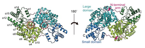 Fig. 1 The homodimeric crystal structure of SpAST.1
Key structural properties of SPAST:
- AAA ATPase hexameric ring structure
- Microtubule binding domain (MTBD)
- Microtubule recognition loop (M-loop)
- Hydrolyzed domain (Walker B motif)
Functions of SPAST
The main function of the spastin protein encoded by the SPAST gene is to cut and reshape the microtubule network of cells. However, it is also involved in a variety of key cellular physiological processes, including vesicle transport, organelle localization and cell division.
| Function | Description |
| Microtubule cutting | Mechanical force is generated by hydrolyzing ATP to specifically cut microtubules and regulate microtubule dynamics and network recombination. |
| Axon Transport support | To clear path obstacles for intracellular vesicles and organelles moving along microtubules and ensure efficient material transport on neuronal axons. |
| Mitotic regulation | Regulate the stability and tension of the spindle microtubules during cell division to ensure the correct separation of chromosomes. |
| Maintenance of endoplasmic reticulum morphology | The three-dimensional tubular network structure of the endoplasmic reticulum is shaped by cutting microtubules to influence its function. |
| Stress response | It is activated under cellular stress conditions (such as damage) and assists cells in adaptation and repair through large-scale reorganization of micronetworks. |
Unlike motor proteins such as kinesin that "walk along the microtubules", spastin acts as a "microtubule cutter" by hydrolyzing ATP at specific points to directly break the microtubule precursor, which enables it to play a unique regulatory role in maintaining the plasticity of the cytoskeleton.
Applications of SPAST and SPAST Antibody in Literature
1. Jeong, Soo Yeon, Hyeonseok Jin, and Jeong Ho Chang. "Crystal structure of L-aspartate aminotransferase from Schizosaccharomyces pombe." PLoS One 14.8 (2019): e0221975. https://doi.org/10.1371/journal.pone.0221975
In this paper, by determining the crystal structure of Schizoyeast L-aspartate aminotransferase 2.1A, it was found that it regulates the catalytic mechanism through conformational changes in the N-terminal arm. Based on the characteristics of the N-terminal arm, the related enzymes were classified into eight subcategories, revealing the structural evolution relationship of pyridoxal phosphate dependent enzymes.
2. Mohan, Neha, et al. "Therapeutic strategies for mutant SPAST-based hereditary spastic paraplegia." Brain sciences 11.8 (2021): 1081. https://doi.org/10.3390/brainsci11081081
The article indicates that SPAST gene mutation is the main cause of hereditary spastic paraplegia, which leads to the loss of spastin protein function and the acquisition of toxic functions. This article explores therapeutic strategies targeting these two pathogenic mechanisms to alleviate the symptoms of patients with SPG4-type HSP.
3. Elert-Dobkowska, Ewelina, et al. "SPAST intragenic CNVs lead to hereditary spastic paraplegia via a haploinsufficiency mechanism." International journal of molecular sciences 25.9 (2024): 5008. https://doi.org/10.3390/ijms25095008
The article indicates that through the analysis of 69 SPG4 patients, it was found that the repeated Alu sequences within the SPAST gene are highly prone to cause micro-rearrangement mutations. The research successfully located multiple gene breakpoints and revealed that non-allelic homologous recombination is its formation mechanism.
4. Nan, Haitian, et al. "A p. Arg499His mutation in SPAST is associated with infantile-onset complicated spastic paraplegia: a case report and review of the literature." BMC neurology 21.1 (2021): 439. https://doi.org/10.1186/s12883-021-02478-0
The article indicates that the p.Arg499His mutation in the SPAST gene is closely related to the complex hereditary spastic paraplegia that occurs in infancy. The literature review concludes that this mutant phenotype can be classified into three categories, and the new mutation rate is high. This discovery expands its clinical spectrum.
5. Verriello, Lorenzo, et al. "Amplifying the spectrum of SPAST gene mutations." Acta Bio Medica: Atenei Parmensis 92.Suppl 1 (2021): e2021220. https://doi.org/10.23750/abm.v92iS1.11608
The article indicates that a novel variation of the SPAST gene was discovered in a pure-phenotypic HSP family in Italy. The 3T microdeletion in exon 13 leads to the deletion of leucine at position 508 of the AAA domain, which is highly conserved. It is speculated that this will disrupt protein function and cause disease.
Creative Biolabs: SPAST Antibodies for Research
Creative Biolabs specializes in the production of high-quality SPAST antibodies for research and industrial applications. Our portfolio includes monoclonal antibodies tailored for ELISA, Flow Cytometry, Western blot, immunohistochemistry, and other diagnostic methodologies.
- Custom SPAST Antibody Development: Tailor-made solutions to meet specific research requirements.
- Bulk Production: Large-scale antibody manufacturing for industry partners.
- Technical Support: Expert consultation for protocol optimization and troubleshooting.
- Aliquoting Services: Conveniently sized aliquots for long-term storage and consistent experimental outcomes.
For more details on our SPAST antibodies, custom preparations, or technical support, contact us at email.
Reference
- Jeong, Soo Yeon, Hyeonseok Jin, and Jeong Ho Chang. "Crystal structure of L-aspartate aminotransferase from Schizosaccharomyces pombe." PLoS One 14.8 (2019): e0221975. https://doi.org/10.1371/journal.pone.0221975
Anti-SPAST antibodies
 Loading...


Hot products 
-
Mouse Anti-ALPL Antibody (B4-78) (CBMAB-1009CQ)

-
Mouse Anti-ADAM12 Recombinant Antibody (V2-179752) (CBMAB-A1114-YC)

-
Mouse Anti-BCL6 Recombinant Antibody (CBYY-0442) (CBMAB-0445-YY)

-
Rabbit Anti-ADRA1A Recombinant Antibody (V2-12532) (CBMAB-1022-CN)

-
Mouse Anti-AMACR Recombinant Antibody (CB34A) (CBMAB-CA034LY)

-
Mouse Anti-BMI1 Recombinant Antibody (CBYC-P026) (CBMAB-P0108-YC)

-
Mouse Anti-BSN Recombinant Antibody (219E1) (CBMAB-1228-CN)

-
Human Anti-SARS-CoV-2 Spike Recombinant Antibody (CBC05) (CBMAB-CR005LY)

-
Mouse Anti-CCL18 Recombinant Antibody (64507) (CBMAB-C7910-LY)

-
Rabbit Anti-ABL1 (Phosphorylated Y185) Recombinant Antibody (V2-443434) (PTM-CBMAB-0001YC)

-
Mouse Anti-AAV-5 Recombinant Antibody (V2-503417) (CBMAB-V208-1369-FY)

-
Mouse Anti-ALX1 Recombinant Antibody (96k) (CBMAB-C0616-FY)

-
Mouse Anti-EPO Recombinant Antibody (CBFYR0196) (CBMAB-R0196-FY)

-
Mouse Anti-C5AR1 Recombinant Antibody (R63) (CBMAB-C9553-LY)

-
Human Anti-SARS-CoV-2 Spike Recombinant Antibody (CR3022) (CBMAB-CR014LY)

-
Mouse Anti-C1QC Recombinant Antibody (CBFYC-0600) (CBMAB-C0654-FY)

-
Mouse Anti-ALOX5 Recombinant Antibody (33) (CBMAB-1890CQ)

-
Mouse Anti-AQP2 Recombinant Antibody (G-3) (CBMAB-A3359-YC)

-
Mouse Anti-FTH1 Recombinant Antibody (CBXF-1896) (CBMAB-F3426-CQ)

-
Mouse Anti-ELAVL4 Recombinant Antibody (6B9) (CBMAB-1132-YC)

- AActivation
- AGAgonist
- APApoptosis
- BBlocking
- BABioassay
- BIBioimaging
- CImmunohistochemistry-Frozen Sections
- CIChromatin Immunoprecipitation
- CTCytotoxicity
- CSCostimulation
- DDepletion
- DBDot Blot
- EELISA
- ECELISA(Cap)
- EDELISA(Det)
- ESELISpot
- EMElectron Microscopy
- FFlow Cytometry
- FNFunction Assay
- GSGel Supershift
- IInhibition
- IAEnzyme Immunoassay
- ICImmunocytochemistry
- IDImmunodiffusion
- IEImmunoelectrophoresis
- IFImmunofluorescence
- IGImmunochromatography
- IHImmunohistochemistry
- IMImmunomicroscopy
- IOImmunoassay
- IPImmunoprecipitation
- ISIntracellular Staining for Flow Cytometry
- LALuminex Assay
- LFLateral Flow Immunoassay
- MMicroarray
- MCMass Cytometry/CyTOF
- MDMeDIP
- MSElectrophoretic Mobility Shift Assay
- NNeutralization
- PImmunohistologyp-Paraffin Sections
- PAPeptide Array
- PEPeptide ELISA
- PLProximity Ligation Assay
- RRadioimmunoassay
- SStimulation
- SESandwich ELISA
- SHIn situ hybridization
- TCTissue Culture
- WBWestern Blot
