TGFB1 Antibodies
Background
TGFB1 is a pleiotropic cytokine widely present in vertebrate tissues, which is secreted in the form of a latent complex and then activated in the body. The protein encoded by this gene plays a core role in tissue repair, embryonic development and inflammatory response by regulating processes such as cell proliferation, differentiation, apoptosis and immune response. Its dysfunction is closely related to fibrosis, cancer and autoimmune diseases. TGFB1 was first identified by Michael Sporn's team in 1983 and is the most distinctive member of the TGF-β superfamily. The analysis of its three-dimensional structure and signal transduction pathways has provided a key foundation for the development of targeted therapeutic drugs. The in-depth study of this gene has greatly advanced our understanding of cell signal transduction, tumor microenvironment and immune regulatory mechanisms.
Structure of TGFB1
TGFB1 is a homodimer protein with a molecular weight of approximately 44 kDa. The size of its monomers varies slightly among different species due to the hydrolysis processing of precursor proteins and sequence differences.
| Species | Human | Mouse | Rat | Pig | Bovine |
| Molecular Weight (kDa) | 44.2 | 43.8 | 44.0 | 44.1 | 44.3 |
| Primary Structural Differences | Containing LAP domains, highly conservative | They bind in a different way to their precursors | Slightly different activation state conformation | Similar to the receptor binding region | The sequences of latent associated peptides are slightly different |
This protein is composed of a precursor peptide of 390 amino acids, which is cleaved by protease to produce an active monomer of 112 amino acids. Its spatial structure is centered around a characteristic "cysteine junction" motif, consisting of a rigid framework formed by four antiparallel β -folds, and intramchain disulfide bonds are formed through nine conserved cysteine residues. The C-terminal α-helix is responsible for binding to type I receptors, and the receptor binding groove is composed of β-strand ring regions, which specifically recognize receptors on the cell surface. Latent associated peptide (LAP) maintains proteins in an inactive state through non-covalent interactions, and this regulatory mechanism plays a key role in the TGF-β signaling pathway.
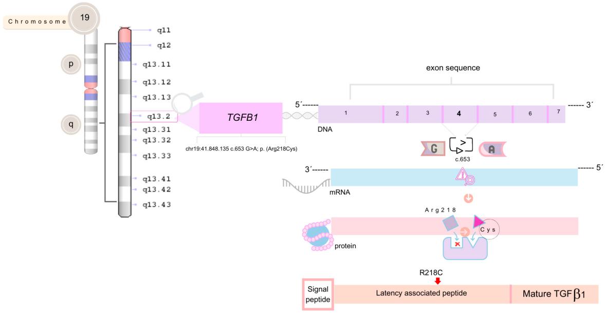 Fig. 1 A Missense Variant (p.Arg218Cys) in the TGFB1 LAP Domain.1
Key structural properties of TGFB1:
- Conservative cysteine junction structure
- Hydrophobic core keep folding conformation growth factors
- Latent associated peptide (LAP) shields active sites through non-covalent interactions
Functions of TGFB1
The main function of the protein encoded by the TGFB1 gene is to regulate cell growth, differentiation and immune response, while also participating in tissue repair and fibrosis processes.
| Function | Description |
| Regulation of cell proliferation | Inhibit the growth of epithelial cells and hematopoietic cells, induce the proliferation of stromal cells, and maintain tissue homeostasis. |
| Immune regulation | Inhibit lymphocyte activation and the production of inflammatory factors, regulate immune tolerance and autoimmune responses. |
| Cell differentiation induction | Promote the differentiation of fibroblasts into myofibroblasts, and participate in wound healing and tissue fibrosis. |
| Regulation of embryonic development | Adjust the mesoderm formation, cell apoptosis and organogenesis, affecting the normal development of embryos. |
| Tumor suppression and promotion | Inhibiting tumor formation in the early stage and promoting tumor metastasis and microenvironment remodeling in the late stage have dual effects. |
The signal activation of TGFB1 follows a dual-pathway pattern of SMAD-dependent and SMad-independent. After binding to the receptor, it forms a phosphorylated Smad complex that enters the nucleus to regulate gene expression. Its dose-effect shows a biphase characteristic: low concentrations inhibit cell growth, while high concentrations promote fibrosis and tumor progression.
Applications of TGFB1 and TGFB1 Antibody in Literature
1. Wang, Cui-zhu, et al. "Comprehensive characterization of TGFB1 across hematological malignancies." Scientific Reports 13.1 (2023): 19107. https://doi.org/10.1038/s41598-023-46552-8
The article indicates that TGFB1 is generally abnormally expressed in hematological malignancies and is associated with a poor prognosis. This gene affects the response to immunotherapy by regulating the tumor immune microenvironment and can serve as a potential target and predictive marker for immunotherapy.
2. Lu, Yingchang, et al. "TGFB1 genetic polymorphisms and coronary heart disease risk: a meta-analysis." BMC medical genetics 13.1 (2012): 39. https://doi.org/10.1186/1471-2350-13-39
The article indicates that variations in the TGFB1 gene are associated with the risk of coronary heart disease. Meta-analysis shows that carriers of the secondary alleles rs1800469 and rs1982073 have a 14% and 18% increased risk of coronary heart disease, respectively, suggesting that the TGFB1 polymorphism is a potential genetic risk factor for CHD.
3. Trugilo, Kleber Paiva, et al. "Haplotype structures and protein levels of TGFB1 in HPV infection and cervical lesion: a case-control study." Cells 12.1 (2022): 84. https://doi.org/10.3390/cells12010084
Research has found that specific variations of the TGFB1 gene (such as c.1347C >T, c.29C>T) and haplotype *3 significantly increase the risk of HPV infection and affect the level of plasma TGF-β1, indicating that they play an important role in the development of cervical lesions.
4. Wodziński, Damian, et al. "Assessment of the TGFB1 gene expression and methylation status of the promoter region in patients with colorectal cancer." Scientific Reports 12.1 (2022): 11488. https://doi.org/10.1038/s41598-022-15599-4
This study shows that the expression level of the TGFB1 gene in colorectal cancer is related to the TNM stage, and higher expression is associated with no vascular invasion and lymphocyte infiltration, suggesting that its down-regulation may promote tumor progression.
5. Szaluś-Jordanow, Olga, et al. "A primary multiple pleomorphic rhabdomyosarcoma of the heart in an adult dog." BMC Veterinary Research 19.1 (2023): 137. https://doi.org/10.1371/journal.pone.0093938
Meta-analysis indicates that the high expression (HP) haplotype of the TGFB1 gene significantly increases the risk of acute rejection in recipients of solid organ transplants, especially heart transplants, while the medium and low expression haplotypes have a lower risk.
Creative Biolabs: TGFB1 Antibodies for Research
Creative Biolabs specializes in the production of high-quality TGFB1 antibodies for research and industrial applications. Our portfolio includes monoclonal antibodies tailored for ELISA, Flow Cytometry, Western blot, immunohistochemistry, and other diagnostic methodologies.
- Custom TGFB1 Antibody Development: Tailor-made solutions to meet specific research requirements.
- Bulk Production: Large-scale antibody manufacturing for industry partners.
- Technical Support: Expert consultation for protocol optimization and troubleshooting.
- Aliquoting Services: Conveniently sized aliquots for long-term storage and consistent experimental outcomes.
For more details on our TGFB1 antibodies, custom preparations, or technical support, contact us at email.
Reference
- Campos, Talyta, et al. "Phenotypic variability in Camurati–Engelmann disease: A case report of a family with the c. 653G> A Pathogenic variant in the TGFB1 Gene." Genes 15.11 (2024): 1354. https://doi.org/10.3390/genes15111354
Anti-TGFB1 antibodies
 Loading...


Hot products 
-
Mouse Anti-ARIH1 Recombinant Antibody (C-7) (CBMAB-A3563-YC)

-
Mouse Anti-CALR Recombinant Antibody (CBFYC-0763) (CBMAB-C0818-FY)

-
Mouse Anti-ACKR3 Recombinant Antibody (V2-261265) (CBMAB-C1023-LY)

-
Mouse Anti-GFP Recombinant Antibody (28) (CBMAB-G3038-LY)

-
Mouse Anti-CCS Recombinant Antibody (CBFYC-1093) (CBMAB-C1150-FY)

-
Mouse Anti-14-3-3 Pan Recombinant Antibody (V2-9272) (CBMAB-1181-LY)

-
Mouse Anti-BACE1 Recombinant Antibody (CBLNB-121) (CBMAB-1180-CN)

-
Mouse Anti-AOC3 Recombinant Antibody (CBYY-0014) (CBMAB-0014-YY)

-
Rat Anti-CD63 Recombinant Antibody (7G4.2E8) (CBMAB-C8725-LY)

-
Mouse Anti-ELAVL4 Recombinant Antibody (6B9) (CBMAB-1132-YC)

-
Mouse Anti-CD24 Recombinant Antibody (ALB9) (CBMAB-0176CQ)

-
Mouse Anti-CRTAM Recombinant Antibody (CBFYC-2235) (CBMAB-C2305-FY)

-
Mouse Anti-ENO2 Recombinant Antibody (85F11) (CBMAB-0276CQ)

-
Mouse Anti-BBS2 Recombinant Antibody (CBYY-0253) (CBMAB-0254-YY)

-
Mouse Anti-EMP3 Recombinant Antibody (CBFYE-0100) (CBMAB-E0207-FY)

-
Rat Anti-C5AR1 Recombinant Antibody (8D6) (CBMAB-C9139-LY)

-
Mouse Anti-ASTN1 Recombinant Antibody (H-9) (CBMAB-1154-CN)

-
Mouse Anti-ALDOA Recombinant Antibody (A2) (CBMAB-A2316-YC)

-
Rabbit Anti-AP2M1 (Phosphorylated T156) Recombinant Antibody (D4F3) (PTM-CBMAB-0610LY)

-
Mouse Anti-CORO1A Recombinant Antibody (4G10) (V2LY-1206-LY806)

- AActivation
- AGAgonist
- APApoptosis
- BBlocking
- BABioassay
- BIBioimaging
- CImmunohistochemistry-Frozen Sections
- CIChromatin Immunoprecipitation
- CTCytotoxicity
- CSCostimulation
- DDepletion
- DBDot Blot
- EELISA
- ECELISA(Cap)
- EDELISA(Det)
- ESELISpot
- EMElectron Microscopy
- FFlow Cytometry
- FNFunction Assay
- GSGel Supershift
- IInhibition
- IAEnzyme Immunoassay
- ICImmunocytochemistry
- IDImmunodiffusion
- IEImmunoelectrophoresis
- IFImmunofluorescence
- IGImmunochromatography
- IHImmunohistochemistry
- IMImmunomicroscopy
- IOImmunoassay
- IPImmunoprecipitation
- ISIntracellular Staining for Flow Cytometry
- LALuminex Assay
- LFLateral Flow Immunoassay
- MMicroarray
- MCMass Cytometry/CyTOF
- MDMeDIP
- MSElectrophoretic Mobility Shift Assay
- NNeutralization
- PImmunohistologyp-Paraffin Sections
- PAPeptide Array
- PEPeptide ELISA
- PLProximity Ligation Assay
- RRadioimmunoassay
- SStimulation
- SESandwich ELISA
- SHIn situ hybridization
- TCTissue Culture
- WBWestern Blot
