TLR3 Antibodies
Background
TLR3 is an important pattern recognition receptor, mainly expressed on the endosomal membrane of immune cells and epithelial cells. The protein encoded by this gene can specifically recognize viral double-stranded RNA, thereby activating the NF-κB and interferon signaling pathways and inducing the occurrence of inflammatory factors and antiviral responses. In the process of host defense against RNA virus (such as influenza virus and norovirus) infections, TLR3 plays a key role by initiating the innate immune response. This gene was discovered in 1997 and is the first receptor in the Toll-like receptor family to have its recognition mechanism clarified. Its unique ligand recognition domain and signal transduction mechanism provide important targets for immunotherapy and vaccine development. Continuous research on TLR3 has deepened people's understanding of innate immune signal transduction, virus-host interactions, and the mechanisms of autoimmune diseases.
Structure of TLR3
TLR3 is a type I transmembrane protein receptor with a molecular weight of approximately 125 kDa. This value may fluctuate slightly among different mammals due to subtle differences in amino acid sequences.
| Species | Human | Mouse | Rat | Bovine | Rhesus monkey |
| Molecular Weight (kDa) | 125.4 | 126.1 | 125.8 | 125.6 | 125.3 |
| Primary Structural Differences | Contains 23 LRR, highly conserved structure of TIR domain | The number of LRS is the same, and the sequence homology is approximately 80% | High similarity to the human TIR domain | Species-specific variation was found in the extracellular domain | More than 90% homology to human sequence |
TLR3 is composed of approximately 904 amino acids, and its extracellular domain forms a horseshoe-shaped structure, responsible for recognizing double-stranded RNA. This receptor is connected to the intracellular TIR domain through a transmembrane region, which initiates downstream signal transduction. The ligand binding site of TLR3 is composed of the LRR module, which can specifically bind to dsRNA, while the TIR domain recruits the adaptor protein TRIF, ultimately activating the expression of interferons and inflammatory factors.
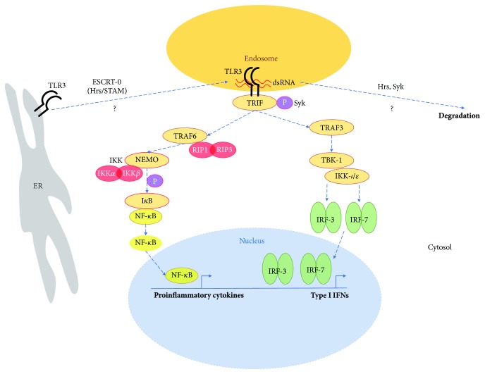 Fig. 1 TLR3 signaling in astrocytes.1
Key structural properties of TLR3:
- Horseshoe-shaped extracellular domains rich in leucine repeat sequences (LRR)
- The transmembrane region is anchored to the cell membrane and connects the extracellular and intracellular domains
- The intracellular Toll/ interleukin-1 receptor (TIR) domain is responsible for signal transduction
- Specifically recognize viral double-stranded RNA and activate immune response pathways
Functions of TLR3
The main function of TLR3 is to recognize viral double-stranded RNA and activate the innate immune response. However, it is also involved in a variety of pathophysiological processes, including the occurrence of autoimmune diseases, the regulation of inflammatory cytokine storms, and in some cases, its impact on tissue regeneration.
| Function | Description |
| Viral Recognition | TLR3 specifically recognizes double-stranded RNA (dsRNA) generated during viral replication and is a key molecule for the body to sense viral invasion. |
| Immune Activation | After recognizing viral RNA, TLR3 activates the NF-κB and IRF signaling pathways, inducing the production of type I interferons and inflammatory factors, and initiating an antiviral immune response. |
| Linking to Adaptive Immunity | By activating dendritic cells, TLR3 promotes antigen presentation, thereby bridging innate immunity and adaptive immunity and activating T-cell responses. |
| Apoptosis Regulation | In some cell types, the activation of TLR3 can induce apoptosis to clear virus-infected cells and prevent the further spread of the virus. |
| Autoimmunity & Disease | In addition to antiviral effects, the abnormal activation of TLR3 is also believed to be related to the pathogenesis of some autoimmune diseases, such as lupus erythematosus. |
The ligand recognition pattern of TLR3 is highly specific, in contrast to the extensive recognition of ssRNA by TLR7/8, which indicates its core role in specifically monitoring the invasion of dsRNA viruses.
Applications of TLR3 and TLR3 Antibody in Literature
1. Wang, Yuru, et al. "TLR3 activation by Clonorchis sinensis infection alleviates the fluke-induced liver fibrosis." PLoS Neglected Tropical Diseases 17.5 (2023): e0011325. https://doi.org/10.1371/journal.pntd.0011325
The article indicates that the absence of TLR3 aggravates liver inflammation and fibrosis caused by Clonoris sinensis infection, while Poly (I:C) alleviates parasite charge and liver fibrosis by activating the TLR3 signaling pathway. TLR3 alleviates fibrosis by inhibiting the p38/ERK pathway and reducing the expression of IL-6 and TNF.
2. Zhang, Xueying, et al. "Extracellular RNAs-TLR3 signaling contributes to cognitive impairment after chronic neuropathic pain in mice." Signal Transduction and Targeted Therapy 8.1 (2023): 292. https://doi.org/10.1038/s41392-023-01543-z
The article indicates that chronic neuralgia leads to an increase in extracellular RNA (especially dsRNA) in the hippocampus, activates TLR3 and promotes inflammation and neuronal apoptosis, resulting in cognitive impairment. Inhibiting TLR3 or using dsRNA/TLR3 antagonists can improve cognitive function, alleviate neuroinflammation and synaptic damage.
3. Wang, Ben‐Gang, De‐Hui Yi, and Yong‐Feng Liu. "TLR3 gene polymorphisms in cancer: a systematic review and meta‐analysis." Cancer Communications 34.3 (2015): 1-13. https://doi.org/10.1186/s40880-015-0020-z
The article indicates that the polymorphism of the TLR3 gene is associated with cancer risk. Meta-analysis shows that the rs5743312 variant genotype significantly increases the overall cancer risk; The polymorphisms of rs3775290 and rs3775291 are also associated with an increased risk in Asian populations and specific subgroups.
4. Lin, Li, et al. "TLR3 Knockdown Attenuates Pressure‐Induced Neuronal Damage In Vitro." Journal of Cellular and Molecular Medicine 28.23 (2024): e70276. https://doi.org/10.1111/jcmm.70276
The article indicates that in spinal cord injury, the upregulation of TLR3 expression exacerbates stress-induced motor neuron injury and apoptosis. Inhibition of TLR3 can alleviate damage, promote mitochondrial autophagy and improve mitochondrial function through the TLR3/IRF3 and TLF3/NF-κB pathways.
5. Ko, Kwang Hyun, et al. "A novel defined TLR3 agonist as an effective vaccine adjuvant." Frontiers in Immunology 14 (2023): 1075291. https://doi.org/10.3389/fimmu.2023.1075291
The article indicates that the novel TLR3 agonist NexaVant (NVT) is a structurally stable and homogeneous dsRNA molecule, which can effectively activate TLR3 and downstream signaling pathways, promote dendritic cell maturation and Th1-type immune responses, and has good safety. It is a promising vaccine adjuvant.
Creative Biolabs: TLR3 Antibodies for Research
Creative Biolabs specializes in the production of high-quality TLR3 antibodies for research and industrial applications. Our portfolio includes monoclonal antibodies tailored for ELISA, Flow Cytometry, Western blot, immunohistochemistry, and other diagnostic methodologies.
- Custom TLR3 Antibody Development: Tailor-made solutions to meet specific research requirements.
- Bulk Production: Large-scale antibody manufacturing for industry partners.
- Technical Support: Expert consultation for protocol optimization and troubleshooting.
- Aliquoting Services: Conveniently sized aliquots for long-term storage and consistent experimental outcomes.
For more details on our TLR3 antibodies, custom preparations, or technical support, contact us at email.
Reference
- Mielcarska, Matylda B., et al. "Syk and Hrs Regulate TLR3‐Mediated Antiviral Response in Murine Astrocytes." Oxidative Medicine and Cellular Longevity 2019.1 (2019): 6927380. https://doi.org/10.1155/2019/6927380
Anti-TLR3 antibodies
 Loading...


Hot products 
-
Mouse Anti-DMPK Recombinant Antibody (CBYCD-324) (CBMAB-D1200-YC)

-
Mouse Anti-GDF5 Recombinant Antibody (1F4) (CBMAB-G2740-LY)

-
Mouse Anti-dsDNA Recombinant Antibody (22) (CBMAB-AP1954LY)

-
Mouse Anti-AMH Recombinant Antibody (5/6) (CBMAB-A2527-YC)

-
Mouse Anti-GIPC2 Recombinant Antibody (10) (CBMAB-G0476-LY)

-
Mouse Anti-CD247 Recombinant Antibody (6B10.2) (CBMAB-C1583-YY)

-
Mouse Anti-ATP1B1 Recombinant Antibody (E4) (CBMAB-0463-LY)

-
Mouse Anti-GFAP Recombinant Antibody (20) (CBMAB-G2914-LY)

-
Mouse Anti-DLG1 Monolconal Antibody (4F3) (CBMAB-0225-CN)

-
Mouse Anti-CD59 Recombinant Antibody (CBXC-2097) (CBMAB-C4421-CQ)

-
Mouse Anti-GGT1 Recombinant Antibody (1F9) (CBMAB-G3273-LY)

-
Mouse Anti-AMIGO2 Recombinant Antibody (CBYY-C0756) (CBMAB-C2192-YY)

-
Mouse Anti-ACE2 Recombinant Antibody (V2-179293) (CBMAB-A0566-YC)

-
Mouse Anti-14-3-3 Pan Recombinant Antibody (V2-9272) (CBMAB-1181-LY)

-
Mouse Anti-ARID1B Recombinant Antibody (KMN1) (CBMAB-A3546-YC)

-
Rat Anti-ABCC11 Recombinant Antibody (V2-179001) (CBMAB-A0236-YC)

-
Rabbit Anti-B2M Recombinant Antibody (CBYY-0059) (CBMAB-0059-YY)

-
Mouse Anti-CDK7 Recombinant Antibody (CBYY-C1783) (CBMAB-C3221-YY)

-
Mouse Anti-F11R Recombinant Antibody (402) (CBMAB-0026-WJ)

-
Mouse Anti-Acetyl-α-Tubulin (Lys40) Recombinant Antibody (V2-623485) (CBMAB-CP2897-LY)

- AActivation
- AGAgonist
- APApoptosis
- BBlocking
- BABioassay
- BIBioimaging
- CImmunohistochemistry-Frozen Sections
- CIChromatin Immunoprecipitation
- CTCytotoxicity
- CSCostimulation
- DDepletion
- DBDot Blot
- EELISA
- ECELISA(Cap)
- EDELISA(Det)
- ESELISpot
- EMElectron Microscopy
- FFlow Cytometry
- FNFunction Assay
- GSGel Supershift
- IInhibition
- IAEnzyme Immunoassay
- ICImmunocytochemistry
- IDImmunodiffusion
- IEImmunoelectrophoresis
- IFImmunofluorescence
- IGImmunochromatography
- IHImmunohistochemistry
- IMImmunomicroscopy
- IOImmunoassay
- IPImmunoprecipitation
- ISIntracellular Staining for Flow Cytometry
- LALuminex Assay
- LFLateral Flow Immunoassay
- MMicroarray
- MCMass Cytometry/CyTOF
- MDMeDIP
- MSElectrophoretic Mobility Shift Assay
- NNeutralization
- PImmunohistologyp-Paraffin Sections
- PAPeptide Array
- PEPeptide ELISA
- PLProximity Ligation Assay
- RRadioimmunoassay
- SStimulation
- SESandwich ELISA
- SHIn situ hybridization
- TCTissue Culture
- WBWestern Blot
