USP7 Antibodies
Background
USP7 is a deubiquitinating enzyme mainly composed of ubiquitin-specific proteases, widely present in the nucleoplasm of eukaryotic cells. This enzyme specifically recognizes and cleaves the ubiquitin chains on the substrate proteins, thereby regulating the stability and function of various key proteins, involving important biological processes such as cell cycle, DNA repair, and epigenetic modification. In cancer and neurodegenerative disease research, USP7 has received significant attention due to its regulatory role in tumor suppressor factors such as p53 and PTEN. Since its first identification in 1997, USP7 has become one of the most representative members of the deubiquitinating enzyme family. The study of its structural characteristics and mechanism of action has greatly promoted the development of drugs targeting the ubiquitin-proteasome system, providing a theoretical basis for targeted treatment of related diseases.
Structure of USP7
USP7 is a large-molecular-weight deubiquitinating enzyme. Its molecular weight varies due to post-translational modifications and splicing variants. The molecular weight of the full-length human USP7 protein is approximately 130-135 kDa. This enzyme belongs to the cysteine protease family and contains multiple functional domains. The most characteristic ones are the N-terminal TRAF-like domain and the C-terminal catalytic domain. The TRAF-like domain is responsible for recognizing and binding to various substrate proteins, such as p53, MDM2, etc.; while the catalytic domain contains a conserved "cysteine-histidine-aspartic acid" catalytic triad, which is responsible for hydrolyzing the ubiquitin molecular chains attached to the substrate protein, thereby regulating the stability and function of the target protein.
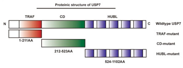 Fig. 1 Schematic illustration of the protein structure of USP7.1
Key structural properties of USP7:
- Typical ubiquitin-specific protease folding structure
- The central catalytic domain contains a Cys-His-Asp/Asn triad active site.
- The N-terminal TRAF-like domain mediates substrate recognition and protein interaction
- The C-terminal extension region is involved in regulating enzyme activity and cell localization
- Catalytic activity is highly dependent on activity at a cystine a nucleophilic attack
Functions of USP7
The core function of the USP7 gene is to regulate the stability and activity of key proteins within cells. It achieves this by specifically cutting the ubiquitin chains on the substrate proteins. This gene is involved in maintaining cellular homeostasis, responding to DNA damage, and regulating various important physiological and pathological processes such as epigenetic state regulation.
| Function | Description |
| Protein Deubiquitination | As a deubiquitinating enzyme, it hydrolyzes the ubiquitin chains on the substrate proteins (such as p53, MDM2, PTEN, etc.), preventing them from being degraded by the proteasome and thereby regulating their stability. |
| Maintenance of genomic stability | By stabilizing DNA repair-related proteins (such as CHK1, RNF168, etc.), it participates in the DNA damage response and repair process. |
| Cell Cycle Regulation | Regulates the stability of multiple cell cycle checkpoint proteins, influencing cell proliferation and division processes. |
| Epigenetic Regulation | By deubiquitinating histones or chromatin-modifying enzymes (such as DNMT1, EZH2, etc.), it affects the gene expression pattern. |
| Dual Role of Tumor Suppression and Promotion | Depending on the cellular context and substrate selection, it can either stabilize tumor suppressor proteins such as p53 to exert protective effects, or promote the accumulation of oncogenic proteins such as MDM2 to facilitate tumor development. |
Unlike the single-function oxygen-storing protein myoglobin, USP7 plays a complex and crucial regulatory role in the cellular signaling network through dynamic and selective deubiquitination. Its dysfunction is closely related to various diseases such as cancer and neurodegenerative disorders.
Applications of USP7 and USP7 Antibody in Literature
1. Ni, Wenkai, et al. "USP7 mediates pathological hepatic de novo lipogenesis through promoting stabilization and transcription of ZNF638." Cell Death & Disease 11.10 (2020): 843. https://doi.org/10.1038/s41419-020-03075-8
Studies have shown that the deubiquitinating enzyme USP7 stabilizes ZNF638, activates the AKT/mTORC1/S6K pathway and forms a nuclear complex, thereby promoting the regulation of SREBP1C on fatty acid synthase genes, and driving the generation of liver lipids. Inhibiting the USP7/ZNF638 axis can significantly alleviate the progression of the mouse fatty liver model, providing a new target for the treatment of related liver diseases.
2. Liu, Huai, et al. "USP7 inhibits the progression of nasopharyngeal carcinoma via promoting SPLUNC1-mediated M1 macrophage polarization through TRIM24." Cell death & disease 14.12 (2023): 852. https://doi.org/10.1038/s41419-023-06368-w
Studies have shown that the deubiquitinating enzyme USP7 stabilizes the TRIM24 protein, promotes the expression of SPLUNC1 mediated by STAT3, thereby driving macrophages to polarize towards the M1 phenotype and activating the JAK/STAT pathway. M1 macrophages can inhibit the proliferation and migration of nasopharyngeal cancer cells, and the USP7/TRIM24/SPLUNC1 axis provides a new target for anti-tumor immunotherapy.
3. Dai, Xiaomeng, et al. "USP7 targeting modulates anti-tumor immune response by reprogramming Tumor-associated Macrophages in Lung Cancer." Theranostics 10.20 (2020): 9332. https://doi.org/10.7150/thno.47137
Studies have shown that inhibiting USP7, which is highly expressed in M2-type macrophages, can promote the reversion of these cells to the M1 phenotype by activating the p38 MAPK pathway, thereby enhancing the function of CD8+ T cells. The combination of USP7 inhibitors and PD-1 blockade can synergistically inhibit the growth of lung cancer, providing a new strategy for tumor immunotherapy.
4. Keshri, Swati, et al. "USP7 protects TFEB from proteasome-mediated degradation." Cell Reports 43.11 (2024). https://doi.org/10.1016/j.celrep.2024.114872
Studies have confirmed that the deubiquitinating enzyme USP7 removes the K48-linked polyubiquitin chains of the TFEB protein, preventing it from being degraded by the proteasome, thereby stabilizing the levels of TFEB in the nucleus and cytoplasm, and maintaining the functions of autophagy and lysosome biogenesis in the cells. This provides a new basis for targeting the USP7-TFEB axis to treat cancer and other metabolic-related diseases.
5. Manea, Teodora, et al. "USP7 controls NGN3 stability and pancreatic endocrine lineage development." Nature Communications 14.1 (2023): 2457. https://doi.org/10.1038/s41467-023-38146-9
Studies have shown that the deubiquitinating enzyme USP7 can bind to the transcription factor NGN3 and remove its ubiquitination modification, thereby stabilizing the protein level of NGN3. This mechanism is crucial for driving the differentiation of pancreatic endocrine precursor cells and the formation of pancreatic islet β cells, providing a new target for diabetes cell therapy.
Creative Biolabs: USP7 Antibodies for Research
Creative Biolabs specializes in the production of high-quality USP7 antibodies for research and industrial applications. Our portfolio includes monoclonal and polyclonal antibodies tailored for ELISA, Flow Cytometry, Western blot, immunohistochemistry, and other diagnostic methodologies.
- Custom USP7 Antibody Development: Tailor-made solutions to meet specific research requirements.
- Bulk Production: Large-scale antibody manufacturing for industry partners.
- Technical Support: Expert consultation for protocol optimization and troubleshooting.
- Aliquoting Services: Conveniently sized aliquots for long-term storage and consistent experimental outcomes.
For more details on our USP7 antibodies, custom preparations, or technical support, contact us at email.
Reference
- Ni, Wenkai, et al. "USP7 mediates pathological hepatic de novo lipogenesis through promoting stabilization and transcription of ZNF638." Cell Death & Disease 11.10 (2020): 843. Distributed under Open Access license CC BY 4.0. Cropped from the original figure. https://doi.org/10.1038/s41419-020-03075-8
Anti-USP7 antibodies
 Loading...


Hot products 
-
Mouse Anti-AQP2 Recombinant Antibody (G-3) (CBMAB-A3359-YC)

-
Mouse Anti-CD24 Recombinant Antibody (2Q1282) (CBMAB-C1624-CN)

-
Mouse Anti-BLNK Recombinant Antibody (CBYY-0623) (CBMAB-0626-YY)

-
Mouse Anti-CD1C Recombinant Antibody (L161) (CBMAB-C2173-CQ)

-
Mouse Anti-CASP8 Recombinant Antibody (CBYY-C0987) (CBMAB-C2424-YY)

-
Mouse Anti-CASP7 Recombinant Antibody (10-01-62) (CBMAB-C2005-LY)

-
Mouse Anti-BBS2 Recombinant Antibody (CBYY-0253) (CBMAB-0254-YY)

-
Mouse Anti-AMOT Recombinant Antibody (CBYC-A564) (CBMAB-A2552-YC)

-
Mouse Anti-CORO1A Recombinant Antibody (4G10) (V2LY-1206-LY806)

-
Mouse Anti-ADV Recombinant Antibody (V2-503423) (CBMAB-V208-1364-FY)

-
Mouse Anti-CFL1 (Phospho-Ser3) Recombinant Antibody (CBFYC-1770) (CBMAB-C1832-FY)

-
Mouse Anti-CFL1 Recombinant Antibody (CBFYC-1771) (CBMAB-C1833-FY)

-
Mouse Anti-FOXA3 Recombinant Antibody (2A9) (CBMAB-0377-YC)

-
Mouse Anti-CCT6A/B Recombinant Antibody (CBXC-0168) (CBMAB-C5570-CQ)

-
Mouse Anti-ACTB Recombinant Antibody (V2-179553) (CBMAB-A0870-YC)

-
Mouse Anti-AQP2 Recombinant Antibody (E-2) (CBMAB-A3358-YC)

-
Mouse Anti-AMIGO2 Recombinant Antibody (CBYY-C0756) (CBMAB-C2192-YY)

-
Mouse Anti-FeLV g27 Recombinant Antibody (1) (CBMAB-V208-1714-FY)

-
Mouse Anti-AFDN Recombinant Antibody (V2-58751) (CBMAB-L0408-YJ)

-
Rabbit Anti-ENO2 Recombinant Antibody (BA0013) (CBMAB-0272CQ)

- AActivation
- AGAgonist
- APApoptosis
- BBlocking
- BABioassay
- BIBioimaging
- CImmunohistochemistry-Frozen Sections
- CIChromatin Immunoprecipitation
- CTCytotoxicity
- CSCostimulation
- DDepletion
- DBDot Blot
- EELISA
- ECELISA(Cap)
- EDELISA(Det)
- ESELISpot
- EMElectron Microscopy
- FFlow Cytometry
- FNFunction Assay
- GSGel Supershift
- IInhibition
- IAEnzyme Immunoassay
- ICImmunocytochemistry
- IDImmunodiffusion
- IEImmunoelectrophoresis
- IFImmunofluorescence
- IGImmunochromatography
- IHImmunohistochemistry
- IMImmunomicroscopy
- IOImmunoassay
- IPImmunoprecipitation
- ISIntracellular Staining for Flow Cytometry
- LALuminex Assay
- LFLateral Flow Immunoassay
- MMicroarray
- MCMass Cytometry/CyTOF
- MDMeDIP
- MSElectrophoretic Mobility Shift Assay
- NNeutralization
- PImmunohistologyp-Paraffin Sections
- PAPeptide Array
- PEPeptide ELISA
- PLProximity Ligation Assay
- RRadioimmunoassay
- SStimulation
- SESandwich ELISA
- SHIn situ hybridization
- TCTissue Culture
- WBWestern Blot
