ACTB Antibodies
Background
The ACTB gene is widely present in eukaryotes, and the β -actin it encodes is a core component of cytoskeletal microfilaments. This protein forms a microfilament network through polymerization, which not only maintains the cell's morphology and mechanical strength but also participates in key life activities such as cell movement, cytoplasmic division, and intracellular substance transport. During the process of cell migration, the dynamic recombination of β -actin directly regulates the formation and contraction mechanisms of pseudopodia. This characteristic makes ACTB an important model molecule in cell biology research. Since its identification in the 1970s, this gene has been widely applied in phylogenetic analysis and gene expression research due to its high conservation and fundamental biological significance. Its promoter region is often used as an internal reference benchmark in molecular biology experiments, providing a basic theoretical framework for the study of cell structure and function as well as disease mechanisms.
Structure of ACTB
The molecular weight of β -actin encoded by the ACTB gene is approximately 42 kDa, and this value is highly conserved among different species. Its amino acid sequence contains approximately 375 residues, forming a spherical structure with ATP/ actin binding fissures. This protein polymerizes to form microfilaments, which constitute the core components of the cytoskeleton.
| Species | Human | Mouse | Bovine | Chicken | Zebrafish |
| Molecular Weight (kDa) | 42 | 42 | 42 | 42 | 42 |
| Primary Structural Differences | Containing N-terminal acetylation modification | 99% homologous to humans | Conserved ATP binding sites | The typical bivalent cation combined with domain | Retain the core aggregation functional area |
The spherical domain of this protein is composed of four subdomains, among which the fissures formed by subdomain 1 and subdomain 3 are responsible for ATP binding. This characteristic structure is highly stable during the evolution of eukaryotes. The acidic amino acid region at its N-terminal provides an interaction interface for other actin binding proteins, and this structural characteristic supports its core functions in key life activities such as cell movement and cytoplasmic division.
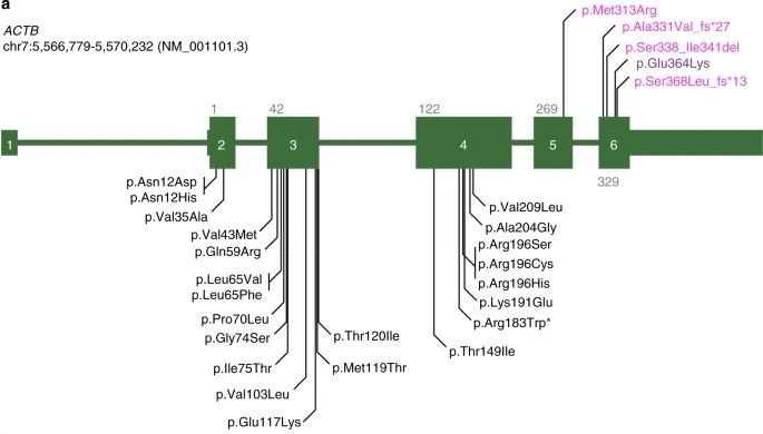 Fig. 1 Schematic representations of ACTB mutations.1
Key structural properties of ACTB:
- Conformation globular monomer contains four structure subdomains
- Conserved ATP/ divalent cation binding fissures
- Highly exposed to N end with C end as protein interaction interface
Functions of ACTB
The β -actin encoded by the ACTB gene mainly functions to form the cytoskeleton and maintain cell morphology, while also participating in a variety of key physiological activities:
| Function | Description |
| Maintenance of cell morphology | By aggregating to form a microfilament network, a cellular mechanical scaffold is constructed to resist external pressure. |
| Regulation of cell movement | Drive the extension and contraction of pseudopodia in cells, providing a structural basis for cell migration. |
| Cytoplasmic division is carried out | At the end of cell division, a contraction ring is formed, completing the physical separation of the two daughter cells. |
| Intracellular substance transport | As a molecular orbital, it supports the directional transport of vesicles and organelles driven by myosin. |
| Regulation of gene expression | Involved in chromatin remodeling and transcription complex assembly via intramuscular actin. |
β -actin achieves functional transformation through the dynamic balance between G-actin monomers and F-actin fibers. Its polymerization process is synergistic regulated by ATP hydrolysis and coproteins. This structural plasticity makes it a core hub connecting cellular physical properties and biological signal transduction.
Applications of ACTB and ACTB Antibody in Literature
1. Dai, Shangzhi, Huijun Wang, and Zhimiao Lin. "ACTB Mutations Analysis and Genotype–Phenotype Correlation in Becker's Nevus." Biomedicines 9.12 (2021): 1879. https://doi.org/10.3390/biomedicines9121879
In this study, it was found that among 20 Chinese patients with Baker's nevus, the majority had a c.C439A/T somatic mutation in the ACTB gene, and the mutation was not limited to the rector pili muscle. More than 20% of the patients were not detected with this mutation, suggesting genetic heterogeneity. Genotype-phenotypic analysis revealed that larger lesions such as the upper trunk and upper limbs were more prone to ACTB mutations.
2. Cuvertino, Sara, et al. "ACTB loss-of-function mutations result in a pleiotropic developmental disorder." The American Journal of Human Genetics 101.6 (2017): 1021-1033. https://doi.org/10.1016/j.ajhg.2017.11.006
This study is the first to confirm that loss-of-function mutations in the ACTB gene can lead to a novel syndrome. The patient presents with developmental delay, intellectual disability, and malformations of the heart/kidneys, etc. Mechanistically, an insufficient single dose of ACTB can alter cell morphology, migration and proliferation, and affect gene expression, thereby damaging organ development.
3. Liu, Chunlan, et al. "ACTB methylation in blood as a potential marker for the pre-clinical detection of stroke: a prospective nested case-control study." Frontiers in neuroscience 15 (2021): 644943. https://doi.org/10.3389/fnins.2021.644943
This study is the first to find that hypomethylation of the ACTB gene in the blood is significantly associated with a short-term (especially within 2 years) risk of stroke. The lower the methylation level, the higher the risk. This indicates that ACTB methylation can serve as a novel biomarker for early risk warning of stroke.
4. Jialie, et al. "The association between ACTB methylation in peripheral blood and coronary heart disease in a case-control study." Frontiers in cardiovascular medicine 9 (2022): 972566. https://doi.org/10.3389/fcvm.2022.972566
This study is the first to find that hypermethylation of the ACTB gene in the blood is significantly associated with the risk of coronary heart disease in Chinese people, especially in men and patients with heart failure. The combined detection of multiple CpG loci can effectively distinguish coronary heart disease, suggesting that it can be used as a new risk assessment marker.
5. Li, Mengxia, et al. "ACTB variants confer the genetic susceptibility to diabetic kidney disease in a Han Chinese population." Frontiers in Genetics 10 (2019): 663. https://doi.org/10.3389/fgene.2019.00663
In this study, it was found that the common variants rs852426 and rs2966449 of the ACTB gene in the Han population with type 2 diabetes were significantly associated with the genetic susceptibility to diabetic nephropathy (DKD). Among them, rs852426 is particularly significantly associated with the risk of DKD in people over 70 years old, providing evidence for the role of ACTB in DKD.
Creative Biolabs: ACTB Antibodies for Research
Creative Biolabs specializes in the production of high-quality ACTB antibodies for research and industrial applications. Our portfolio includes monoclonal antibodies tailored for ELISA, Flow Cytometry, Western blot, immunohistochemistry, and other diagnostic methodologies.
- Custom ACTB Antibody Development: Tailor-made solutions to meet specific research requirements.
- Bulk Production: Large-scale antibody manufacturing for industry partners.
- Technical Support: Expert consultation for protocol optimization and troubleshooting.
- Aliquoting Services: Conveniently sized aliquots for long-term storage and consistent experimental outcomes.
For more details on our ACTB antibodies, custom preparations, or technical support, contact us at email.
Reference
- Latham, Sharissa L., et al. "Variants in exons 5 and 6 of ACTB cause syndromic thrombocytopenia." Nature communications 9.1 (2018): 4250. https://doi.org/10.1038/s41467-018-06713-0
Anti-ACTB antibodies
 Loading...


Hot products 
-
Mouse Anti-BRCA2 Recombinant Antibody (CBYY-1728) (CBMAB-2077-YY)

-
Mouse Anti-C1QC Recombinant Antibody (CBFYC-0600) (CBMAB-C0654-FY)

-
Mouse Anti-BLNK Recombinant Antibody (CBYY-0623) (CBMAB-0626-YY)

-
Mouse Anti-AP4E1 Recombinant Antibody (32) (CBMAB-A2996-YC)

-
Mouse Anti-ACLY Recombinant Antibody (V2-179314) (CBMAB-A0610-YC)

-
Mouse Anti-CASP7 Recombinant Antibody (10-01-62) (CBMAB-C2005-LY)

-
Mouse Anti-CCN2 Recombinant Antibody (CBFYC-2383) (CBMAB-C2456-FY)

-
Mouse Anti-ADRB2 Recombinant Antibody (V2-180026) (CBMAB-A1420-YC)

-
Rabbit Anti-ATF4 Recombinant Antibody (D4B8) (CBMAB-A3872-YC)

-
Mouse Anti-AAV8 Recombinant Antibody (V2-634028) (CBMAB-AP022LY)

-
Mouse Anti-ACTB Recombinant Antibody (V2-179553) (CBMAB-A0870-YC)

-
Mouse Anti-ACKR3 Recombinant Antibody (V2-261265) (CBMAB-C1023-LY)

-
Mouse Anti-CD1C Recombinant Antibody (L161) (CBMAB-C2173-CQ)

-
Mouse Anti-BRCA2 Recombinant Antibody (CBYY-0790) (CBMAB-0793-YY)

-
Mouse Anti-AAV9 Recombinant Antibody (V2-634029) (CBMAB-AP023LY)

-
Mouse Anti-CEMIP Recombinant Antibody (3C12) (CBMAB-K0296-LY)

-
Mouse Anti-AHCYL1 Recombinant Antibody (V2-180270) (CBMAB-A1703-YC)

-
Mouse Anti-dsDNA Recombinant Antibody (22) (CBMAB-AP1954LY)

-
Mouse Anti-CA9 Recombinant Antibody (CBXC-2079) (CBMAB-C0131-CQ)

-
Mouse Anti-DHFR Recombinant Antibody (D0821) (CBMAB-D0821-YC)

- AActivation
- AGAgonist
- APApoptosis
- BBlocking
- BABioassay
- BIBioimaging
- CImmunohistochemistry-Frozen Sections
- CIChromatin Immunoprecipitation
- CTCytotoxicity
- CSCostimulation
- DDepletion
- DBDot Blot
- EELISA
- ECELISA(Cap)
- EDELISA(Det)
- ESELISpot
- EMElectron Microscopy
- FFlow Cytometry
- FNFunction Assay
- GSGel Supershift
- IInhibition
- IAEnzyme Immunoassay
- ICImmunocytochemistry
- IDImmunodiffusion
- IEImmunoelectrophoresis
- IFImmunofluorescence
- IGImmunochromatography
- IHImmunohistochemistry
- IMImmunomicroscopy
- IOImmunoassay
- IPImmunoprecipitation
- ISIntracellular Staining for Flow Cytometry
- LALuminex Assay
- LFLateral Flow Immunoassay
- MMicroarray
- MCMass Cytometry/CyTOF
- MDMeDIP
- MSElectrophoretic Mobility Shift Assay
- NNeutralization
- PImmunohistologyp-Paraffin Sections
- PAPeptide Array
- PEPeptide ELISA
- PLProximity Ligation Assay
- RRadioimmunoassay
- SStimulation
- SESandwich ELISA
- SHIn situ hybridization
- TCTissue Culture
- WBWestern Blot
