AMOT Antibodies
Background
AMOT is a scaffold protein, mainly expressed in endothelial cells and epithelial cells of vertebrates. The protein encoded by this gene plays a key role in embryonic development and the maintenance of tissue homeostasis by participating in processes such as cell polarity establishment, cell migration and angiogenesis. It is particularly worth noting that AMOT can interact with the core components of the Hippo signaling pathway, thereby regulating organ size and tumor suppressive function. Scientists first identified the AMOT gene in 2003. Subsequent research has found that its abnormal expression is closely related to various cancers and vascular diseases. As an important component of cell junction complexes, AMOT has become a hot topic in cell biology and oncology research due to its unique molecular mechanism, providing a new perspective for understanding tissue morphogenesis and cancer metastasis.
Structure of AMOT
Myoglobin is a relatively small protein with a molecular weight of approximately 16.7 kDa. This weight may slightly vary between species due to minor differences in amino acid sequence.
| Species | Human | Mouse | Rat | Zebrafish |
| Molecular Weight (kDa) | ~100 (AMOT p130) | ~98 (AMOT p130) | ~100 (AMOT p130) | ~85 (homologous gene) |
| Primary Structural Differences | Containing PPxY motif, PDZ binding domain | Highly conservative and functionally similar | Highly homologous to human AMOT | Retain the regulatory function of the Hippo pathway |
The AMOT protein is composed of multiple functional domains, including the n-terminal coiled-coil domain, the intermediate PDZ binding domain, and the c-terminal PPxY motif. These structures enable it to interact with various signaling molecules, such as YAP/TAZ and LATS kinases. Its tertiary structure forms a stable scaffold, participates in the assembly of cell junction complexes, and plays a key role in cell polarity, migration and angiogenesis. The subcellular localization of AMOT (such as in the cell membrane or cytoplasm) is regulated by phosphorylation, which affects its functional activity.
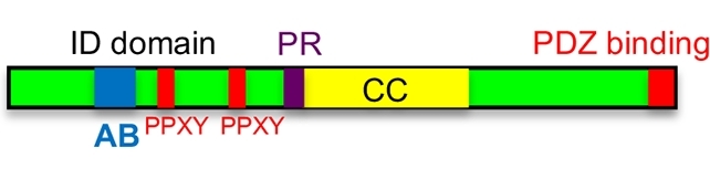 Fig. 1 Domain architecture of Amot protein. 1
Key structural properties of AMOT:
- Multi-domain scaffold protein
- Hippo pathway regulatory core
- Phosphorylation-dependent functional regulation
- Cell junction localization
Functions of AMOT
The core function of the AMOT gene is to regulate cell polarity and migration, and it is also involved in a variety of physiological and pathological processes, including angiogenesis, tumor suppression and mechanical signal transduction.
| Function | Description |
| Establishment of cell polarity | By connecting the cell membrane with the cytoskeleton, it regulates the apical and basal polarity of epithelial cells and endothelial cells, maintaining the function of the tissue barrier. |
| Hippo pathway regulation | As a key inhibitor of YAP/TAZ, it inhibits excessive cell proliferation and tumorigenesis through LATS kinase-dependent phosphorylation. |
| Angiogenesis regulation | By integrating VEGF and Angiopoietin signals, endothelial cell migration and vascular lumen formation are controlled, influencing embryonic development and wound repair. |
| Mechanical force induction | In response to changes in extracellular matrix hardness, mechanical signals are transmitted through cell junction complexes (such as tight junctions) to regulate cell morphology and migration. |
| Tumor metastasis inhibition | In various cancers (such as breast cancer and liver cancer), the absence of AMOT expression can lead to the activation of YAP, promoting tumor invasion and metastasis. |
The functional exertion of AMOT depends on its subcellular localization (such as at cell junctions or in the cytoplasm) and phosphorylation status. Its mechanism of action is more complex than that of traditional scaffold proteins and involves cross-regulation of multiple signaling pathways.
Applications of AMOT and AMOT Antibody in Literature
1. Rojek, Katarzyna O., et al. "Amot and Yap1 regulate neuronal dendritic tree complexity and locomotor coordination in mice." PLoS biology 17.5 (2019): e3000253.https://doi.org/10.1371/journal.pbio.3000253
Research has found that angioactin (Amot) and Hippo pathway co-activator Yap1 jointly promote the dendrite development of hippocampal neurons and Purkinje cells by regulating the phosphorylation of S6 kinase (rather than TEAD transcription factors), and their absence can lead to abnormal cerebellar morphology and motor coordination disorders.
2. Rheinemann, Lara, et al. "Interactions between AMOT PPxY motifs and NEDD4L WW domains function in HIV-1 release." Journal of Biological Chemistry 297.2 (2021). https://doi.org/10.1016/j.jbc.2021.100975
Research has found that the HIV virus binds with high affinity to the WW3 domain of NEDD4L through the PPxY1 motif of the AMOT protein, promoting the formation and release of the viral envelope. Among them, ionic and hydrophobic interactions are the key mechanisms.
3. Jiang, Lai, et al. "Cell size regulates human endoderm specification through actomyosin-dependent AMOT-YAP signaling." Stem Cell Reports 19.8 (2024): 1137-1155.https://doi.org/10.1016/j.stemcr.2024.07.001
Research has found that when human stem cells differentiate into the endoderm, the cell volume decreases. Hypertonic pressure promotes AMOT nuclear translocation by activating actibulin and inhibits YAP activity, thereby enhancing endoderm differentiation.
4. Heller, Brigitte, et al. "Amot recognizes a juxtanuclear endocytic recycling compartment via a novel lipid binding domain." Journal of Biological Chemistry 285.16 (2010): 12308-12320. https://doi.org/10.1074/jbc.m109.096230
Research has found that the polar protein Amot specifically recognizes phosphatidylinositol monophosphate and cholesterol through a novel lipid-binding domain, mediating the translocation of the Patj/Mupp1 complex from the plasma membrane to the systemic circulation compartment, thereby regulating cell polarity.
5. Zhu, Guoqing, et al. "12-O-Tetradecanoylphorbol-13-acetate (TPA) is anti-tumorigenic in liver cancer cells via inhibiting YAP through AMOT." Scientific reports 7.1 (2017): 44940.https://doi.org/10.1038/srep44940
Research has found that TPA promotes the translocation of YAP from the nucleus to the cytoplasm through an AmOT-dependent mechanism, blocking its transcriptional activity and thereby inhibiting the malignant phenotype of liver cancer cells. The absence of AMOT will eliminate the anti-tumor effect of TPA, indicating that the AMOT-YAP axis is the key pathway for TPA to exert its tumor suppression function.
Creative Biolabs: AMOT Antibodies for Research
Creative Biolabs specializes in the production of high-quality AMOT antibodies for research and industrial applications. Our portfolio includes monoclonal antibodies tailored for ELISA, Flow Cytometry, Western blot, immunohistochemistry, and other diagnostic methodologies.
- Custom AMOT Antibody Development: Tailor-made solutions to meet specific research requirements.
- Bulk Production: Large-scale antibody manufacturing for industry partners.
- Technical Support: Expert consultation for protocol optimization and troubleshooting.
- Aliquoting Services: Conveniently sized aliquots for long-term storage and consistent experimental outcomes.
For more details on our AMOT antibodies, custom preparations, or technical support, contact us at email.
Reference
- Rojek, Katarzyna O., et al. "Amot and Yap1 regulate neuronal dendritic tree complexity and locomotor coordination in mice." PLoS biology 17.5 (2019): e3000253.https://doi.org/10.1371/journal.pbio.3000253
Anti-AMOT antibodies
 Loading...


Hot products 
-
Mouse Anti-ALB Recombinant Antibody (V2-180650) (CBMAB-A2186-YC)

-
Mouse Anti-ANXA7 Recombinant Antibody (A-1) (CBMAB-A2941-YC)

-
Mouse Anti-APC Recombinant Antibody (CBYC-A661) (CBMAB-A3036-YC)

-
Mouse Anti-CRTAM Recombinant Antibody (CBFYC-2235) (CBMAB-C2305-FY)

-
Mouse Anti-ATP1B1 Recombinant Antibody (E4) (CBMAB-0463-LY)

-
Mouse Anti-BANF1 Recombinant Antibody (3F10-4G12) (CBMAB-A0707-LY)

-
Mouse Anti-APOE Recombinant Antibody (A1) (CBMAB-0078CQ)

-
Mouse Anti-CCDC6 Recombinant Antibody (CBXC-0106) (CBMAB-C5397-CQ)

-
Mouse Anti-C5B-9 Recombinant Antibody (CBFYA-0216) (CBMAB-X0304-FY)

-
Mouse Anti-HTLV-1 gp46 Recombinant Antibody (CBMW-H1006) (CBMAB-V208-1154-FY)

-
Mouse Anti-ATM Recombinant Antibody (2C1) (CBMAB-A3970-YC)

-
Mouse Anti-APOA1 Monoclonal Antibody (CBFYR0637) (CBMAB-R0637-FY)

-
Mouse Anti-APCS Recombinant Antibody (CBYC-A663) (CBMAB-A3054-YC)

-
Mouse Anti-4-Hydroxynonenal Recombinant Antibody (V2-502280) (CBMAB-C1055-CN)

-
Mouse Anti-CDK7 Recombinant Antibody (CBYY-C1783) (CBMAB-C3221-YY)

-
Mouse Anti-CFL1 (Phospho-Ser3) Recombinant Antibody (CBFYC-1770) (CBMAB-C1832-FY)

-
Mouse Anti-CD33 Recombinant Antibody (6C5/2) (CBMAB-C8126-LY)

-
Mouse Anti-ADGRL2 Recombinant Antibody (V2-58519) (CBMAB-L0166-YJ)

-
Mouse Anti-CCNH Recombinant Antibody (CBFYC-1054) (CBMAB-C1111-FY)

-
Mouse Anti-DMD Recombinant Antibody (D1190) (CBMAB-D1190-YC)

- AActivation
- AGAgonist
- APApoptosis
- BBlocking
- BABioassay
- BIBioimaging
- CImmunohistochemistry-Frozen Sections
- CIChromatin Immunoprecipitation
- CTCytotoxicity
- CSCostimulation
- DDepletion
- DBDot Blot
- EELISA
- ECELISA(Cap)
- EDELISA(Det)
- ESELISpot
- EMElectron Microscopy
- FFlow Cytometry
- FNFunction Assay
- GSGel Supershift
- IInhibition
- IAEnzyme Immunoassay
- ICImmunocytochemistry
- IDImmunodiffusion
- IEImmunoelectrophoresis
- IFImmunofluorescence
- IGImmunochromatography
- IHImmunohistochemistry
- IMImmunomicroscopy
- IOImmunoassay
- IPImmunoprecipitation
- ISIntracellular Staining for Flow Cytometry
- LALuminex Assay
- LFLateral Flow Immunoassay
- MMicroarray
- MCMass Cytometry/CyTOF
- MDMeDIP
- MSElectrophoretic Mobility Shift Assay
- NNeutralization
- PImmunohistologyp-Paraffin Sections
- PAPeptide Array
- PEPeptide ELISA
- PLProximity Ligation Assay
- RRadioimmunoassay
- SStimulation
- SESandwich ELISA
- SHIn situ hybridization
- TCTissue Culture
- WBWestern Blot
