BTLA Antibodies
Background
The BTLA gene is located in the 3q13.2 region of the human chromosome and encodes B and T lymphocyte attenuation factors. It is an immune checkpoint protein mainly expressed on the surface of lymphocytes. This protein regulates the activation threshold of immune cells by binding to HVEM, a member of the tumor necrosis factor receptor superfamily, and transmits inhibitory signals to prevent excessive immune responses and maintain immune homeostasis. Its functional defects are closely related to autoimmune diseases, chronic infections and tumor immune escape. Since it was identified as an immunosuppressive receptor in 2003, it has become an important research target in the field of tumor immunotherapy. The discovery of this gene and the study of its mechanism not only deepen people's understanding of the immune tolerance regulatory network, but also provide a key theoretical basis for the development of new immunotherapies.
Structure of BTLA
BTLA (B and T lymphocyte attenuation factor) is a transmembrane glycoprotein with a molecular weight of approximately 28-32 kDa. Its precise molecular weight varies slightly due to glycosylation modification and species differences.
| Species | Human | Mouse | Rat |
| Molecular Weight (kDa) | ~28-32 | ~26-30 | ~27-31 |
| Primary Structural Differences | Contains two immunoglobulin-like domains, a transmembrane region and an intracellular ITIM motif | Highly homologous to human structure and functionally conserved | With humans and mice have similar domain of protein structure |
This protein is composed of 289 amino acids and belongs to the immunoglobulin superfamily. Its primary structure folds to form two extracellular immunoglobulin-mutable (IgV) domains, constituting the key interface for binding to the ligand HVEM. The secondary structure is mainly composed of β -folds, forming the core framework of immunoglobulin-like folds. The key functional domains include: the extracellular region is responsible for recognizing ligands; Transmembrane region Moreover, the cytoplasmic tail region contains an immune receptor tyrosine inhibitory motif (ITIM). After phosphorylation, this motif can recruit SHP-1/SHP-2 phosphatases, thereby conducting inhibitory signals and regulating lymphocyte activity.
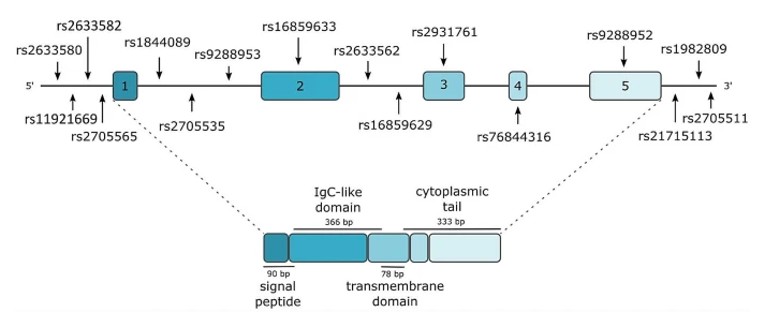 Fig. 1 Structure of the BTLA gene and the BTLA protein along with the location of the studied BTLA gene variants.1
Key structural properties of BTLA:
- Immunoglobulin-like folding structure
- Hydrophobic core and ligand binding interface
- Intracellular signal domains containing ITIM motifs
Functions of BTLA
The core function of the protein encoded by the BTLA gene is to transmit inhibitory signals in the immune system and maintain autoimmune tolerance. However, it also plays a role in various pathophysiological processes, including tumor immune escape and the occurrence of autoimmune diseases.
| Function | Description |
| Immune regulation | Inhibitory signals are transmitted through intracellular ITIM motifs to down-regulate the activation and proliferation of lymphocytes (T/B cells), preventing excessive immune responses. |
| Maintain tolerance | Involved in the establishment and maintenance of immune tolerance in the thymus and peripheral tissues, preventing attacks against autoantigens. |
| Tumor immune escape | In the tumor microenvironment, its signaling pathways are often utilized by cancer cells to suppress anti-tumor immune responses and promote tumor growth. |
| Infection and chronic inflammation | Under chronic viral infection and inflammatory conditions, its persistent high expression may lead to the exhaustion of T cell function and weaken the ability to clear pathogens. |
| Associated with autoimmune diseases | Genetic polymorphisms or functional deficiencies are associated with an increased risk of autoimmune diseases such as rheumatoid arthritis and systemic lupus erythematosus. |
Contrary to the synergistic stimulating effect usually formed by activated immune receptors (such as CD28), the inhibitory signal of BTLA is threshain-regulated. It does not completely shut down the immune response but sets a higher activation threshold to ensure that lymphocytes are fully activated only when exposed to strong antigenic stimulation. This delicate negative feedback regulation mechanism is crucial for balancing effective immune defense and avoiding autoimmune damage.
Applications of BTLA and BTLA Antibody in Literature
1. Yu, Xueping, et al. "BTLA contributes to acute-on-chronic liver failure infection and mortality through CD4+ T-cell exhaustion." Nature Communications 15.1 (2024): 1835. https://doi.org/10.1038/s41467-024-46047-8
The article indicates that in hepatitis B-related acute-on-chronic liver failure, the level of BTLA is upregulated through the IL-6/TNF pathway, inhibits the function of CD4+T cells through the PI3K-Akt pathway, and promotes infection. Blocking BTLA can improve the survival rate and bacterial clearance of mice. This study provides a new target for treatment.
2. Ning, Zhaochen, Keyan Liu, and Huabao Xiong. "Roles of BTLA in immunity and immune disorders." Frontiers in Immunology 12 (2021): 654960. https://doi.org/10.3389/fimmu.2021.654960
The article indicates that BTLA is a key immunosuppressive co-signaling molecule that widely inhibits lymphocyte activity by binding to the ligand HVEM. It plays a significant role in various pathological processes such as tumors, autoimmune diseases, and infections, and is a potential target for immunotherapy.
3. Andrzejczak, Anna, and Lidia Karabon. "BTLA biology in cancer: from bench discoveries to clinical potentials." Biomarker Research 12.1 (2024): 8. https://doi.org/10.1186/s40364-024-00556-2
The article indicates that BTLA, as a key immune checkpoint, negatively regulates immunity through interaction with HVEM. In cancer, the upregulation of BTLA expression can suppress anti-tumor immunity, leading to a poor prognosis. Therapies targeting BTLA have shown potential to enhance anti-tumor immunity in preclinical studies, presenting a new hope for cancer treatment.
4. Demerlé, Clemence, Laurent Gorvel, and Daniel Olive. "BTLA-HVEM couple in health and diseases: insights for immunotherapy in lung cancer." Frontiers in oncology 11 (2021): 682007. https://doi.org/10.3389/fonc.2021.682007
The article indicates that BTLA-HVEM is a new target for immunotherapy of lung cancer. It is similar to PD-1 and can inhibit lymphocyte activity. Its expression is related to tumor progression, especially showing significant potential in PD-L1-negative patients and is expected to become a new generation of immunotherapy.
5. Yu, Xueping, et al. "BTLA/HVEM signaling: milestones in research and role in chronic hepatitis B virus infection." Frontiers in immunology 10 (2019): 617. https://doi.org/10.3389/fimmu.2019.00617
The article indicates that BTLA is an immunosuppressive receptor similar to PD-1 and jointly regulates immunity with the ligand HVEM. It is highly expressed in chronic hepatitis B infection and inhibits T cell function. The specific mechanism remains to be further studied.
Creative Biolabs: BTLA Antibodies for Research
Creative Biolabs specializes in the production of high-quality BTLA antibodies for research and industrial applications. Our portfolio includes monoclonal antibodies tailored for ELISA, Flow Cytometry, Western blot, immunohistochemistry, and other diagnostic methodologies.
- Custom BTLA Antibody Development: Tailor-made solutions to meet specific research requirements.
- Bulk Production: Large-scale antibody manufacturing for industry partners.
- Technical Support: Expert consultation for protocol optimization and troubleshooting.
- Aliquoting Services: Conveniently sized aliquots for long-term storage and consistent experimental outcomes.
For more details on our BTLA antibodies, custom preparations, or technical support, contact us at email.
Reference
- Andrzejczak, Anna, and Lidia Karabon. "BTLA biology in cancer: from bench discoveries to clinical potentials." Biomarker Research 12.1 (2024): 8. https://doi.org/10.1186/s40364-024-00556-2
Anti-BTLA antibodies
 Loading...


Hot products 
-
Mouse Anti-CTNND1 Recombinant Antibody (CBFYC-2414) (CBMAB-C2487-FY)

-
Mouse Anti-BSN Recombinant Antibody (219E1) (CBMAB-1228-CN)

-
Rabbit Anti-B2M Recombinant Antibody (CBYY-0059) (CBMAB-0059-YY)

-
Mouse Anti-C5b-9 Recombinant Antibody (aE11) (CBMAB-AO138LY)

-
Mouse Anti-8-oxoguanine Recombinant Antibody (V2-7719) (CBMAB-1898CQ)

-
Mouse Anti-FAS2 Monoclonal Antibody (1D4) (CBMAB-0071-CN)

-
Mouse Anti-APOE Recombinant Antibody (A1) (CBMAB-0078CQ)

-
Mouse Anti-CAT Recombinant Antibody (724810) (CBMAB-C8431-LY)

-
Mouse Anti-BCL2L1 Recombinant Antibody (H5) (CBMAB-1025CQ)

-
Rat Anti-4-1BB Recombinant Antibody (V2-1558) (CBMAB-0953-LY)

-
Mouse Anti-2C TCR Recombinant Antibody (V2-1556) (CBMAB-0951-LY)

-
Mouse Anti-BrdU Recombinant Antibody (IIB5) (CBMAB-1038CQ)

-
Rabbit Anti-ATF4 Recombinant Antibody (D4B8) (CBMAB-A3872-YC)

-
Rat Anti-FABP3 Recombinant Antibody (CBXF-2299) (CBMAB-F1612-CQ)

-
Mouse Anti-14-3-3 Pan Recombinant Antibody (V2-9272) (CBMAB-1181-LY)

-
Mouse Anti-CARTPT Recombinant Antibody (113612) (CBMAB-C2450-LY)

-
Mouse Anti-CTCF Recombinant Antibody (CBFYC-2371) (CBMAB-C2443-FY)

-
Mouse Anti-AMH Recombinant Antibody (5/6) (CBMAB-A2527-YC)

-
Mouse Anti-HTLV-1 gp46 Recombinant Antibody (CBMW-H1006) (CBMAB-V208-1154-FY)

-
Rat Anti-ADGRE4 Recombinant Antibody (V2-160163) (CBMAB-F0011-CQ)

- AActivation
- AGAgonist
- APApoptosis
- BBlocking
- BABioassay
- BIBioimaging
- CImmunohistochemistry-Frozen Sections
- CIChromatin Immunoprecipitation
- CTCytotoxicity
- CSCostimulation
- DDepletion
- DBDot Blot
- EELISA
- ECELISA(Cap)
- EDELISA(Det)
- ESELISpot
- EMElectron Microscopy
- FFlow Cytometry
- FNFunction Assay
- GSGel Supershift
- IInhibition
- IAEnzyme Immunoassay
- ICImmunocytochemistry
- IDImmunodiffusion
- IEImmunoelectrophoresis
- IFImmunofluorescence
- IGImmunochromatography
- IHImmunohistochemistry
- IMImmunomicroscopy
- IOImmunoassay
- IPImmunoprecipitation
- ISIntracellular Staining for Flow Cytometry
- LALuminex Assay
- LFLateral Flow Immunoassay
- MMicroarray
- MCMass Cytometry/CyTOF
- MDMeDIP
- MSElectrophoretic Mobility Shift Assay
- NNeutralization
- PImmunohistologyp-Paraffin Sections
- PAPeptide Array
- PEPeptide ELISA
- PLProximity Ligation Assay
- RRadioimmunoassay
- SStimulation
- SESandwich ELISA
- SHIn situ hybridization
- TCTissue Culture
- WBWestern Blot
