CD1a Antibodies
Background
The CD1a gene encodes a glycoprotein mainly present on the surface of antigen-presenting cells. This protein belongs to the CD1 family and is responsible for presenting lipid antigens to T cells to initiate an immune response. It is highly expressed in thymic cortical epithelial cells and Langerhans cells, playing a crucial role in skin immune surveillance and defense against pathogen invasion. This gene was first identified in the early 1980s. Its uniqueness lies in its ability to present hydrophobic antigens different from classical MHC molecules, thus filling the gap in the immune recognition system. The research on the structure and function of CD1a has greatly advanced the understanding of lipid antigen presentation mechanisms in immunology and provided new molecular targets for infectious diseases, autoimmune diseases and tumor immunotherapy.
Structure of CD1a
CD1a is a type I transmembrane glycoprotein with a molecular weight of approximately 33-35 kDa. This molecular weight shows certain differences among different species, mainly due to the degree of glycosylation modification of its extracellular domain and subtle amino acid sequence variations.
| Species | Human | Mouse |
| Molecular Weight (kDa) | ~33-35 | ~35 |
| Primary Structural Differences | It is mainly expressed in Langerhans cells and cortical thymus epithelial cells | There are interspecies differences in the ability to present specific lipid antigens due to different expression patterns |
This protein is structurally composed of three extracellular domains (α1-α3), a transmembrane region and a short intracellular tail. Its α1 and α2 domains fold to form hydrophobic antigen-binding grooves. This structural feature enables it to bind and present various lipid and glycolipid antigens, which is the core basis of its immune function.
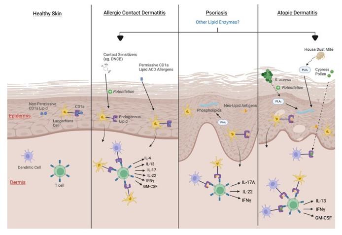 Fig. 1 Postulated mechanisms of CD1a involvement in inflammatory skin disease pathogenesis.1
Key structural properties of CD1a:
- Unique lipid antigen-binding groove structure
- Mainly made up of two alpha helix hydrophobic binding pocket
- MHC-I folding but with non-polymorphic characteristics
- Transmembrane domains are anchored to the surface of antigen-presenting cells
Functions of CD1a
The main function of the CD1a gene-encoded protein is to present lipid antigens to T cells to initiate adaptive immune responses. In addition, it is also involved in various immune regulatory processes, including the establishment of autoimmune tolerance and the regulation of inflammatory responses.
| Function | Description |
| Lipid Antigen presentation | It captures and presents endogenous or pathogen-derived lipid antigens through its antigen-binding grooves, activating CD1A-restricted T cells (such as NKT cells). |
| Immune surveillance | It is mainly expressed on the surface of Langerhans cells and acts as an immune sentinel of the skin barrier, recognizing lipid components related to pathogens. |
| Thymus selection | Positive selection involved in the development of T cells within the thymus is crucial for the formation of self-lipid antigen tolerance. |
| Inflammatory Regulation | Under certain pathological conditions, abnormal lipid antigen presentation mediated by CD1a can drive the occurrence and development of inflammatory skin diseases (such as psoriasis). |
| Tumor Immunology | It is expressed in some tumor microenvironments such as melanoma, and its function is related to the anti-tumor immune response or immune escape mechanism. |
Unlike the classical MHC molecules that present polypeptide antigens, the CD1a protein specifically presents hydrophobic lipid antigens. This characteristic fills the gap in the immune system's recognition of non-protein antigens and is the uniqueness of its immunological function.
Applications of CD1a and CD1a Antibody in Literature
1. Ye, John H., Yi-Ling Chen, and Graham Ogg. "CD1a and skin T cells: a pathway for therapeutic intervention." Clinical and Experimental Dermatology 49.5 (2024): 450-458. https://doi.org/10.1093/ced/llad460
The article indicates that CD1a is a molecule continuously expressed on skin Langerhans cells, responsible for presenting lipid antigens to T cells and participating in immune surveillance. Studies have shown that the dysregulation of T-cell responses mediated by it is related to the pathogenesis of inflammatory skin diseases such as psoriasis, atopic dermatitis and allergic contact dermatitis.
2. Hardman, Clare S., et al. "CD1a promotes systemic manifestations of skin inflammation." Nature Communications 13.1 (2022): 7535. https://doi.org/10.1038/s41467-022-35071-1
Research has found that the CD1a molecule on the surface of skin Langerhans cells can link local skin inflammation (such as TLR7 agonists or MC903 induction) with systemic immune responses (splenomegaly, T cell expansion and cytokine elevation), and its specific blocking antibodies can effectively inhibit this process.
3. Maeda, Sachiko, et al. "Analysis of CD1a-Positive Monocyte-Derived Cells in the Regional Lymph Nodes of Patients with Gallbladder Cancer." International Journal of Molecular Sciences 25.23 (2024): 12763. https://doi.org/10.3390/ijms252312763
This study of 70 cases of gallbladder cancer found that CD1A-positive dendritic cell infiltration in regional lymph nodes was associated with poor prognosis and lymph node metastasis, but it was not an independent prognostic factor. The infiltration of CD1a cells in the primary tumor lesion has a more significant impact on the surgical outcome.
4. Gulic, Tamara, et al. "Human Decidual CD1a+ Dendritic Cells Undergo Functional Maturation Program Mediated by Gp96." International journal of molecular sciences 24.3 (2023): 2278. https://doi.org/10.3390/ijms24032278
This study shows that the heat shock protein gp96 can dose-dependentially bind to the CD91 and TLR4 receptors of decidual CD1a+ dendritic cells, promoting their maturation and increasing the expression of interferon -γ and IL-15, which may enhance the Th1-type immune response at the maternal-fetal interface and affect the maintenance of pregnancy.
5. Mitchell, Jenée, and George Kannourakis. "Does CD1a expression influence T cell function in patients with langerhans cell histiocytosis?." Frontiers in Immunology 12 (2021): 773598. https://doi.org/10.3389/fimmu.2021.773598
This article explores the possibility that the key lesion marker CD1a+ LCH cells in Langerhans cell histiocytosis (LCH) may be recognized by T cells and thereby participate in the pathogenesis of the disease, and points out that targeting this immune interaction is a potential therapeutic direction.
Creative Biolabs: CD1a Antibodies for Research
Creative Biolabs specializes in the production of high-quality CD1a antibodies for research and industrial applications. Our portfolio includes monoclonal antibodies tailored for ELISA, Flow Cytometry, Western blot, immunohistochemistry, and other diagnostic methodologies.
- Custom CD1a Antibody Development: Tailor-made solutions to meet specific research requirements.
- Bulk Production: Large-scale antibody manufacturing for industry partners.
- Technical Support: Expert consultation for protocol optimization and troubleshooting.
- Aliquoting Services: Conveniently sized aliquots for long-term storage and consistent experimental outcomes.
For more details on our CD1a antibodies, custom preparations, or technical support, contact us at email.
Reference
- YaYe, John H., Yi-Ling Chen, and Graham Ogg. "CD1a and skin T cells: a pathway for therapeutic intervention." Clinical and Experimental Dermatology 49.5 (2024): 450-458. https://doi.org/10.1093/ced/llad460
Anti-CD1a antibodies
 Loading...


Hot products 
-
Mouse Anti-APCS Recombinant Antibody (CBYC-A663) (CBMAB-A3054-YC)

-
Mouse Anti-CD1C Recombinant Antibody (L161) (CBMAB-C2173-CQ)

-
Mouse Anti-CD46 Recombinant Antibody (CBFYC-0076) (CBMAB-C0085-FY)

-
Mouse Anti-CD24 Recombinant Antibody (ALB9) (CBMAB-0176CQ)

-
Mouse Anti-BIRC5 Recombinant Antibody (6E4) (CBMAB-CP2646-LY)

-
Mouse Anti-ATM Recombinant Antibody (2C1) (CBMAB-A3970-YC)

-
Rabbit Anti-ENO2 Recombinant Antibody (BA0013) (CBMAB-0272CQ)

-
Rat Anti-ADAM10 Recombinant Antibody (V2-179741) (CBMAB-A1103-YC)

-
Rabbit Anti-CBL Recombinant Antibody (D4E10) (CBMAB-CP0149-LY)

-
Mouse Anti-14-3-3 Pan Recombinant Antibody (V2-9272) (CBMAB-1181-LY)

-
Mouse Anti-AMH Recombinant Antibody (5/6) (CBMAB-A2527-YC)

-
Mouse Anti-FLI1 Recombinant Antibody (CBXF-0733) (CBMAB-F0435-CQ)

-
Mouse Anti-ACTN4 Recombinant Antibody (V2-6075) (CBMAB-0020CQ)

-
Mouse Anti-Acetyl-α-Tubulin (Lys40) Recombinant Antibody (V2-623485) (CBMAB-CP2897-LY)

-
Mouse Anti-ADAM12 Recombinant Antibody (V2-179752) (CBMAB-A1114-YC)

-
Mouse Anti-BZLF1 Recombinant Antibody (BZ.1) (CBMAB-AP705LY)

-
Human Anti-SARS-CoV-2 Spike Recombinant Antibody (CR3022) (CBMAB-CR014LY)

-
Mouse Anti-COL1A2 Recombinant Antibody (CF108) (V2LY-1206-LY626)

-
Mouse Anti-ESR1 Recombinant Antibody (Y31) (CBMAB-1208-YC)

-
Mouse Anti-CD2AP Recombinant Antibody (BR083) (CBMAB-BR083LY)

- AActivation
- AGAgonist
- APApoptosis
- BBlocking
- BABioassay
- BIBioimaging
- CImmunohistochemistry-Frozen Sections
- CIChromatin Immunoprecipitation
- CTCytotoxicity
- CSCostimulation
- DDepletion
- DBDot Blot
- EELISA
- ECELISA(Cap)
- EDELISA(Det)
- ESELISpot
- EMElectron Microscopy
- FFlow Cytometry
- FNFunction Assay
- GSGel Supershift
- IInhibition
- IAEnzyme Immunoassay
- ICImmunocytochemistry
- IDImmunodiffusion
- IEImmunoelectrophoresis
- IFImmunofluorescence
- IGImmunochromatography
- IHImmunohistochemistry
- IMImmunomicroscopy
- IOImmunoassay
- IPImmunoprecipitation
- ISIntracellular Staining for Flow Cytometry
- LALuminex Assay
- LFLateral Flow Immunoassay
- MMicroarray
- MCMass Cytometry/CyTOF
- MDMeDIP
- MSElectrophoretic Mobility Shift Assay
- NNeutralization
- PImmunohistologyp-Paraffin Sections
- PAPeptide Array
- PEPeptide ELISA
- PLProximity Ligation Assay
- RRadioimmunoassay
- SStimulation
- SESandwich ELISA
- SHIn situ hybridization
- TCTissue Culture
- WBWestern Blot
