CDS2 Antibodies
Background
CDS2, as a key member of the cytidine diphosphate diacylglycerol synthase family, is mainly responsible for catalyzing phosphatidylserine into CDP-diacylglycerol, which is an important precursor for the biosynthesis of phosphatidylinositol and cardiolipin. This gene is widely expressed in mammalian tissues, and plays a core role in maintaining the metabolic homeostasis of phosphatidylinositol. Studies have found that the absence of CDS2 leads to defects in phosphatidylinositol synthesis, thereby affecting cell signal transduction and calcium homeostasis regulation. In the field of tumor biology, CDS2 has a synthetic lethal relationship with CDS1, providing a potential therapeutic target for tumors with low expression of CDS1. Additionally, CDS2 is also regulated by p53 and SIRT6 in a synergistic manner, participating in the maintenance of lipid metabolic balance.
Structure of CDS2
The molecular weight of CDS2 protein is approximately 50 kDa and it is highly conserved among different species. This enzyme contains about 400 amino acids and is located in the endoplasmic reticulum. Its core structure includes a catalytic domain and a transmembrane region. The catalytic domain is responsible for converting phosphatidylserine to CDP-diacylglycerol, while the transmembrane region anchors it to the membrane. When the phospholipase C signal is activated, CDS2 regulates cell signal transduction by maintaining the level of phosphatidylinositol. The key amino acid residues in the enzyme's active center ensure the catalytic efficiency.
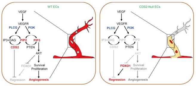 Fig. 1 Working model of VEGFA-triggered vessel regression on CDS2-deficient endothelium.1
Key structural features of CDS2:
- Comprises a catalytic domain and a transmembrane region
- The active center is responsible for catalyzing the formation of CDP-diacylglycerol from phosphatidic acid
- The transmembrane region anchors it to the endoplasmic reticulum membrane
- Key amino acid residues maintain substrate recognition and catalytic efficiency
Functions of CDS2
The main function of CDS2 is to catalyze the synthesis of CDP-diacylglycerol, providing precursors for phosphatidylinositol and cardiolipin. However, it is also involved in various cellular processes, including signal transduction and maintenance of lipid homeostasis.
| Function | Description |
| Phospholipid synthesis | Catalyzes the formation of CDP-diacylglycerol from phosphatidylacylglycerol, which is a key step in the de novo synthesis of phosphatidylinositol. |
| Signal transduction support | When phospholipase C is activated, it maintains the level of phosphatidylinositol through re-synthesis. |
| Lipid homeostasis maintenance | Cooperates with CDS1 to regulate the synthesis of cardiolipin and affects mitochondrial function. |
| Synthetic lethal target | In tumors with low expression of CDS1, the absence of CDS2 induces cell apoptosis, showing therapeutic potential. |
| Calcium homeostasis regulation | Knockout of CDS2 leads to calcium homeostasis imbalance and affects cell signal transduction. |
The enzymatic reaction of CDS2 occurs at the central position of phospholipid metabolism. Its catalytic efficiency directly affects the generation of downstream signaling molecules, and plays a crucial role in maintaining the stability of the cell membrane and energy metabolism.
Applications of CDS2 and CDS2 Antibody in Literature
1. Zhao, Wencao, et al. "Endothelial CDS2 deficiency causes VEGFA-mediated vascular regression and tumor inhibition." Cell research 29.11 (2019): 895-910. https://doi.org/10.1038/s41422-019-0229-5
The research has found that the absence of CDS2 can reverse the function of VEGFA, transforming it from promoting angiogenesis to inducing vascular degeneration. Mechanistically, the stimulation of VEGFA leads to metabolic disorders in phosphatidylinositol, resulting in the deficiency of PIP2/PIP3 and the activation of FOXO1, thereby causing reverse migration of endothelial cells and vascular degeneration.
2. Chan, Pui Ying, et al. "The synthetic lethal interaction between CDS1 and CDS2 is a vulnerability in uveal melanoma and across multiple tumor types." Nature genetics 57.7 (2025): 1672-1683. https://doi.org/10.1038/s41588-025-02222-1
The study found that CDS2 is a genetically-dependent target for uveal melanoma and has a synthetic lethal interaction with CDS1. Knockout of CDS2 disrupts phosphatidylinositol synthesis and promotes cell apoptosis, providing a potential therapeutic strategy for tumors with low expression of CDS1.
3. Collins, Daniel M., et al. "CDS2 expression regulates de novo phosphatidic acid synthesis." Biochemical Journal 481.20 (2024): 1449-1473. https://doi.org/10.1042/BCJ20240456
The study found that after the deletion of CDS2, cells maintained the basal level of phosphatidylinositol by increasing phosphatidylserine de novo synthesis. However, when G protein-coupled receptors were continuously stimulated, the synthesis could not be enhanced, resulting in the loss of PI. Additionally, the knockout of CDS2 also caused calcium homeostasis imbalance.
4. Blunsom, Nicholas J., and Shamshad Cockcroft. "CDP-diacylglycerol synthases (CDS): gateway to phosphatidylinositol and cardiolipin synthesis." Frontiers in cell and developmental biology 8 (2020): 63. https://doi.org/10.3389/fcell.2020.00063
The research has found that the CDS enzyme is a key enzyme for synthesizing phosphatidylinositol and cardiolipin, catalyzing the formation of CDP-diacylglycerol. In mammals, CDS1/2 is located in the endoplasmic reticulum, and CDS2 plays a major role in maintaining the level of phosphatidylinositol during the phospholipase C signaling pathway. Its expression is regulated by transcription.
5. Li, Meiting, et al. "p53 cooperates with SIRT6 to regulate cardiolipin de novo biosynthesis." Cell death & disease 9.10 (2018): 941. https://doi.org/10.1038/s41419-018-0984-0
The study found that p53 and SIRT6 interacted with each other and co-localized at the CDS1/2 promoter in response to palmitic acid stimulation. SIRT6, as a co-activator of p53, recruited RNA polymerase II and enhanced the synthesis of cardiolipin to maintain lipid homeostasis.
Creative Biolabs: CDS2 Antibodies for Research
Creative Biolabs specializes in the production of high-quality CDS2 antibodies for research and industrial applications. Our portfolio includes monoclonal and polyclonal antibodies tailored for ELISA, Flow Cytometry, Western blot, immunohistochemistry, and other diagnostic methodologies.
- Custom CDS2 Antibody Development: Tailor-made solutions to meet specific research requirements.
- Bulk Production: Large-scale antibody manufacturing for industry partners.
- Technical Support: Expert consultation for protocol optimization and troubleshooting.
- Aliquoting Services: Conveniently sized aliquots for long-term storage and consistent experimental outcomes.
For more details on our CDS2 antibodies, custom preparations, or technical support, contact us at email.
Reference
- Zhao, Wencao, et al. "Endothelial CDS2 deficiency causes VEGFA-mediated vascular regression and tumor inhibition." Cell research 29.11 (2019): 895-910. Distributed under Open Access license CC BY 4.0, and cropped from the original figure. https://doi.org/10.1038/s41422-019-0229-5
Anti-CDS2 antibodies
 Loading...


Hot products 
-
Mouse Anti-CASP7 Recombinant Antibody (10-01-62) (CBMAB-C2005-LY)

-
Mouse Anti-CFL1 Recombinant Antibody (CBFYC-1771) (CBMAB-C1833-FY)

-
Mouse Anti-C5AR1 Recombinant Antibody (R63) (CBMAB-C9553-LY)

-
Mouse Anti-FOXL1 Recombinant Antibody (CBXF-0845) (CBMAB-F0462-CQ)

-
Mouse Anti-CD33 Recombinant Antibody (6C5/2) (CBMAB-C8126-LY)

-
Mouse Anti-DES Monoclonal Antibody (440) (CBMAB-AP1857LY)

-
Mouse Anti-EPO Recombinant Antibody (CBFYR0196) (CBMAB-R0196-FY)

-
Mouse Anti-CD33 Recombinant Antibody (P67.6) (CBMAB-C10189-LY)

-
Mouse Anti-BPGM Recombinant Antibody (CBYY-1806) (CBMAB-2155-YY)

-
Mouse Anti-ADGRE2 Recombinant Antibody (V2-261270) (CBMAB-C0813-LY)

-
Mouse Anti-ADV Recombinant Antibody (V2-503423) (CBMAB-V208-1364-FY)

-
Mouse Anti-ENO1 Recombinant Antibody (8G8) (CBMAB-E1329-FY)

-
Mouse Anti-CCL18 Recombinant Antibody (64507) (CBMAB-C7910-LY)

-
Mouse Anti-FOSB Recombinant Antibody (CBXF-3593) (CBMAB-F2522-CQ)

-
Mouse Anti-CDK7 Recombinant Antibody (CBYY-C1783) (CBMAB-C3221-YY)

-
Mouse Anti-ALOX5 Recombinant Antibody (33) (CBMAB-1890CQ)

-
Mouse Anti-BCL6 Recombinant Antibody (CBYY-0442) (CBMAB-0445-YY)

-
Mouse Anti-AQP2 Recombinant Antibody (G-3) (CBMAB-A3359-YC)

-
Mouse Anti-AHCYL1 Recombinant Antibody (V2-180270) (CBMAB-A1703-YC)

-
Mouse Anti-ADRB2 Recombinant Antibody (V2-180026) (CBMAB-A1420-YC)

- AActivation
- AGAgonist
- APApoptosis
- BBlocking
- BABioassay
- BIBioimaging
- CImmunohistochemistry-Frozen Sections
- CIChromatin Immunoprecipitation
- CTCytotoxicity
- CSCostimulation
- DDepletion
- DBDot Blot
- EELISA
- ECELISA(Cap)
- EDELISA(Det)
- ESELISpot
- EMElectron Microscopy
- FFlow Cytometry
- FNFunction Assay
- GSGel Supershift
- IInhibition
- IAEnzyme Immunoassay
- ICImmunocytochemistry
- IDImmunodiffusion
- IEImmunoelectrophoresis
- IFImmunofluorescence
- IGImmunochromatography
- IHImmunohistochemistry
- IMImmunomicroscopy
- IOImmunoassay
- IPImmunoprecipitation
- ISIntracellular Staining for Flow Cytometry
- LALuminex Assay
- LFLateral Flow Immunoassay
- MMicroarray
- MCMass Cytometry/CyTOF
- MDMeDIP
- MSElectrophoretic Mobility Shift Assay
- NNeutralization
- PImmunohistologyp-Paraffin Sections
- PAPeptide Array
- PEPeptide ELISA
- PLProximity Ligation Assay
- RRadioimmunoassay
- SStimulation
- SESandwich ELISA
- SHIn situ hybridization
- TCTissue Culture
- WBWestern Blot
