MTPN Antibodies
Background
The MTPN gene encodes a cytoskeleton-related protein called myponectin, which is widely expressed in vertebrate tissues and has an active function especially in muscle, brain and immune cells. Its encoded protein participates in the maintenance of cell morphology, cytoplasmic division and cell migration processes by regulating actin dynamics, and plays a key role in neuronal development and synaptic formation. This gene was first identified in 1994 when studying the mechanism of cytoskeletal remodeling. Subsequent studies have revealed that it can affect the invasion and metastasis ability of tumor cells by mediating the Rho GTPase signaling pathway. As an important regulatory factor for cell motility and structural remodeling, the study of the molecular mechanism of the MTPN gene provides an important theoretical basis for understanding physiological and pathological processes such as tissue development, immune response and cancer metastasis.
Structure of MTPN
Myotrophin encoded by the MTPN gene is a cellular regulatory protein with a molecular weight of approximately 12.8 kDa. There are slight differences in its molecular weight among different species, mainly due to specific variations in amino acid sequences.
| Species | Human | Mouse | Rat |
| Molecular Weight (kDa) | 12.8 | 12.9 | 12.7 |
| Primary Structural Differences | Contains 3 ANK repeating domains | Very high homology with humans | Key functional domains are highly conserved |
This protein is composed of approximately 125 amino acids, and its core feature is the presence of several ANK repeat sequences, which mediate a wide range of protein-protein interactions. Its three-dimensional structure presents a typical helical - angular - helical fold, forming a highly conserved hydrophobic core, which is crucial for its binding with transcription factors (such as NF-κB) and other signaling molecules, thereby playing a regulatory role in processes such as cell growth, differentiation, and inflammatory responses.
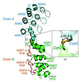 Fig. 1 Overall structure of human V-1(MTPN) in the asymmetric unit.1
Key structural properties of MTPN:
- Contains multiple ANK duplicate domains
- Form a three-dimensional spiral - bundle - spiral folding
- With a highly conserved hydrophobic core area
Functions of MTPN
The core function of the MTPN gene-encoded protein (myotrophic hormone) is to participate in cell growth regulation and signal transduction. In addition, this protein also plays a significant role in various physiological and pathological processes.
| Function | Description |
| Regulation of cell proliferation | By promoting the nuclear translocation of specific transcription factors (such as NF-κB), it positively regulates the growth and proliferation processes of cells. |
| Signal transduction medium | Fill multiple signaling pathways connect molecules inside the cells, involved in regulating cell differentiation and survival. |
| Neuronal development | During the development of the central nervous system, it has a crucial impact on the migration of neurons, the establishment of dendrite morphology and the formation of synapses. |
| Association with cancer progression | Abnormal expression in a variety of tumor tissue, the expression level exists significant correlation with invasion and metastasis of cancer cells ability. |
| Promotion of myocardial hypertrophy | In the cardiac pressure overload model, the upregulation of its expression has been confirmed to be an important driving factor for the pathological occurrence of myocardial hypertrophy. |
This protein interacts with multiple signaling molecules through its ANK repeat domain, and its functional network exhibits a high degree of complexity and context dependence rather than a single linear pathway.
Applications of MTPN and MTPN Antibody in Literature
1. Wang, Yuyao, et al. "The human myotrophin variant attenuates microRNA-Let-7 binding ability but not risk of left ventricular hypertrophy in human essential hypertension." Plos one 10.8 (2015): e0135526. https://doi.org/10.1371/journal.pone.0135526
Studies have found that the rs17168525 variant site on the 3 '-UTR of the myotrophin gene can affect its protein translation: the C allele can promote let-7c binding and inhibit expression, while the T allele weakens this effect. However, this variation has no significant association with hypertensive left ventricular hypertrophy in the Han population.
2. LOOPS, REGULATION BY MYOTROPHIN HAIRPIN. "Nuclear Co-translocation of Myotrophin and p65 Stimulates Myocyte Growth." THE JOURNAL OF BIOLOGICAL CHEMISTRY 283.41 (2008): 27947-27956. https://doi.org/10.1074/JBC.M801210200
Research has found that myotrophin protein co-translocations into the nucleus with p65 through its hairpin ring (especially at the E33A site being crucial), activating NF-κB signaling, thereby stimulating protein synthesis and growth in cardiomyocytes and inducing cardiac hypertrophy. This nuclear translocation is the core mechanism of its hypertrophic function.
3. Zwolak, Adam, et al. "Structural basis for capping protein sequestration by myotrophin (V-1)." Journal of Biological Chemistry 285.33 (2010): 25767-25781. https://doi.org/10.1074/jbc.m110.135848
Research has found that myotrophin binds to the alkaline region of CP through its anchin ring, which is also the site where CP binds to the tips of muscle filaments. The competitive binding of the two leads to the inactivation of CP, preventing it from terminating myofilaments, but myotrophic proteins also cannot dissociate the already terminated CP.
4. Bhattacharya, Nandini, et al. "Binding of myotrophin/V-1 to actin-capping protein: implications for how capping protein binds to the filament barbed end." Journal of Biological Chemistry 281.41 (2006): 31021-31030. https://doi.org/10.1074/jbc.M606278200
Research has found that myptrophin (V-1) directly binds to actin capping protein (CP) with high affinity to form a 1:1 complex, thereby inhibiting the capping activity of CP on the barbed ends of muscle filaments. V-1 binds to the β subunit "tentacle" region of CP through its two anchin rings, and this effect supports the "swing model" of CP binding to myofilaments.
5. Takeda, Shuichi, et al. "Crystal structure of human V-1 in the apo form." Structural Biology and Crystallization Communications 77.1 (2021): 13-21. https://doi.org/10.1107/S2053230X20016829
The article indicates that the structure of myotrophin protein is highly consistent when it is free and bound to CP, with no obvious conformational changes. It can bind to CP through the side chain rearrangement of several residues, thereby widely inhibiting CP activity in the cytoplasm.
Creative Biolabs: MTPN Antibodies for Research
Creative Biolabs specializes in the production of high-quality MTPN antibodies for research and industrial applications. Our portfolio includes monoclonal antibodies tailored for ELISA, Flow Cytometry, Western blot, immunohistochemistry, and other diagnostic methodologies.
- Custom MTPN Antibody Development: Tailor-made solutions to meet specific research requirements.
- Bulk Production: Large-scale antibody manufacturing for industry partners.
- Technical Support: Expert consultation for protocol optimization and troubleshooting.
- Aliquoting Services: Conveniently sized aliquots for long-term storage and consistent experimental outcomes.
For more details on our MTPN antibodies, custom preparations, or technical support, contact us at email.
Reference
- Takeda, Shuichi, et al. "Crystal structure of human V-1 in the apo form." Structural Biology and Crystallization Communications 77.1 (2021): 13-21. https://doi.org/10.1107/S2053230X20016829
Anti-MTPN antibodies
 Loading...


Hot products 
-
Mouse Anti-CAPZB Recombinant Antibody (CBYY-C0944) (CBMAB-C2381-YY)

-
Rabbit Anti-AP2M1 (Phosphorylated T156) Recombinant Antibody (D4F3) (PTM-CBMAB-0610LY)

-
Mouse Anti-DISP2 Monoclonal Antibody (F66A4B1) (CBMAB-1112CQ)

-
Mouse Anti-ASTN1 Recombinant Antibody (H-9) (CBMAB-1154-CN)

-
Mouse Anti-FOXA3 Recombinant Antibody (2A9) (CBMAB-0377-YC)

-
Mouse Anti-ASB9 Recombinant Antibody (1D8) (CBMAB-A0529-LY)

-
Human Anti-SARS-CoV-2 Spike Recombinant Antibody (CBC05) (CBMAB-CR005LY)

-
Mouse Anti-ENO2 Recombinant Antibody (H14) (CBMAB-E1341-FY)

-
Mouse Anti-GGT1 Recombinant Antibody (1F9) (CBMAB-G3273-LY)

-
Rabbit Anti-CBL Recombinant Antibody (D4E10) (CBMAB-CP0149-LY)

-
Mouse Anti-BACE1 Recombinant Antibody (61-3E7) (CBMAB-1183-CN)

-
Mouse Anti-CFL1 Recombinant Antibody (CBFYC-1771) (CBMAB-C1833-FY)

-
Mouse Anti-ADAM12 Recombinant Antibody (V2-179752) (CBMAB-A1114-YC)

-
Human Anti-SARS-CoV-2 Spike Recombinant Antibody (CR3022) (CBMAB-CR014LY)

-
Rabbit Anti-BAD (Phospho-Ser136) Recombinant Antibody (CAP219) (CBMAB-AP536LY)

-
Mouse Anti-14-3-3 Pan Recombinant Antibody (V2-9272) (CBMAB-1181-LY)

-
Mouse Anti-AMACR Recombinant Antibody (CB34A) (CBMAB-CA034LY)

-
Mouse Anti-CHRNA9 Recombinant Antibody (8E4) (CBMAB-C9161-LY)

-
Mouse Anti-ARIH1 Recombinant Antibody (C-7) (CBMAB-A3563-YC)

-
Mouse Anti-AGK Recombinant Antibody (V2-258056) (CBMAB-M0989-FY)

- AActivation
- AGAgonist
- APApoptosis
- BBlocking
- BABioassay
- BIBioimaging
- CImmunohistochemistry-Frozen Sections
- CIChromatin Immunoprecipitation
- CTCytotoxicity
- CSCostimulation
- DDepletion
- DBDot Blot
- EELISA
- ECELISA(Cap)
- EDELISA(Det)
- ESELISpot
- EMElectron Microscopy
- FFlow Cytometry
- FNFunction Assay
- GSGel Supershift
- IInhibition
- IAEnzyme Immunoassay
- ICImmunocytochemistry
- IDImmunodiffusion
- IEImmunoelectrophoresis
- IFImmunofluorescence
- IGImmunochromatography
- IHImmunohistochemistry
- IMImmunomicroscopy
- IOImmunoassay
- IPImmunoprecipitation
- ISIntracellular Staining for Flow Cytometry
- LALuminex Assay
- LFLateral Flow Immunoassay
- MMicroarray
- MCMass Cytometry/CyTOF
- MDMeDIP
- MSElectrophoretic Mobility Shift Assay
- NNeutralization
- PImmunohistologyp-Paraffin Sections
- PAPeptide Array
- PEPeptide ELISA
- PLProximity Ligation Assay
- RRadioimmunoassay
- SStimulation
- SESandwich ELISA
- SHIn situ hybridization
- TCTissue Culture
- WBWestern Blot
