PDK4 Antibodies
Background
PDK4 (Pyruvate Dehydrogenase Kinase 4) is a regulatory enzyme that functions in mitochondria and is mainly present in metabolically active tissues such as the heart, skeletal muscle, and pancreas. This protein inactivates the pyruvate dehydrogenase complex through phosphorylation, thereby inhibiting the conversion of pyruvate to acetyl-CoA, and regulates the metabolic transition between glucose oxidation and fatty acid oxidation. It maintains energy homeostasis under conditions of hunger, diabetes, or high-fat diet. Hibernating animals and mammals in a fasting state rely on PDK4 to adapt to changes in metabolic substrates and ensure the energy supply to vital organs. After its function was systematically elucidated by Sugden and his colleagues in the 1990s, PDK4 has attracted widespread attention due to its crucial role in insulin resistance and metabolic diseases. The determination of its crystal structure provides an important basis for further understanding the regulation of kinase activity and the development of specific inhibitors, and has advanced our understanding of metabolic adaptation, mitochondrial function, and disease mechanisms.
Structure of PDK4
The PDK4 gene encodes the pyruvate dehydrogenase kinase 4 isoform, which has a molecular weight of approximately 46.2 kDa. The molecular weight of this enzyme varies among different species, mainly due to the subtle differences in amino acid sequences.
| Species | Human | Mouse | Rat | Pig | Cow |
| Molecular Weight (kDa) | 46.2 | 46.1 | 46.3 | 46.2 | 46.4 |
| Primary Structural Differences | The kinase domain is highly conserved | About 90% sequence identity with human | A little variation n-terminal sequence exists | High tissue expression specificity | The catalytic domain completely conservative sequence |
The PDK4 gene encodes pyruvate dehydrogenase kinase 4, which contains approximately 411 amino acids. Its three-dimensional structure exhibits the typical fold pattern of mitochondrial protein kinases. This protein consists of a regulatory domain at the N-terminus and a catalytic domain at the C-terminus. The catalytic domain presents a bimodal conformation, with the adenylate binding site located in the gap between the two lobes. The function of PDK4 depends on the conserved amino acid residues in its active center, where Asp282 and Asp318 are coordinated by magnesium ions for the transfer of the phosphate group of ATP, and Cys245 participates in substrate recognition and conformational changes. The secondary structure of this enzyme is characterized by alternating α-helices and β-sheets. The C-terminal domain contains a hydrophobic core that maintains the stability of the catalytic pocket. Compared with other members of the same family, PDK4 has a unique Loop structure near the ATP binding pocket, which affects its selectivity for different nucleotide substrates and regulates the interaction interface with the pyruvate dehydrogenase complex.
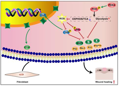 Fig. 1 Concise model of PDK4 in inhibiting HDFs senescence and accelerating wound healing.1
Key structural properties of PDK4:
- Two-domain folding, including the N-terminal regulatory region and the C-terminal catalytic core
- ATP binding pocket is composed of a conserved Gly-rich loop
- Kinase activity depends on the key aspartic acid residues in the catalytic domain
- The unique Loop region structure affects substrate recognition and inhibitor binding
Functions of PDK4
The PDK4 gene encodes pyruvate dehydrogenase kinase 4, which mainly functions to regulate the metabolic conversion between glucose and fatty acids.
| Function | Description |
| Metabolic switch regulation | In the presence of hunger or high-fat diet, it inhibits glucose oxidation and promotes the utilization of fatty acids, maintaining energy homeostasis. |
| Substrate competition regulation | By inactivating pyruvate dehydrogenase, the entry of pyruvate into the tricarboxylic acid cycle is reduced, thereby preserving substrates for gluconeogenesis. |
| Insulin Sensitivity Regulation | When overexpressed in skeletal muscle, it can induce insulin resistance and affect the efficiency of glucose uptake and utilization. |
| Stress Adaptation Response | Upregulated expression under conditions of hypoxia or mitochondrial dysfunction, helping cells adapt to energy metabolism stress. |
| Organ-specific metabolic regulation | Differentially expressed in the heart, liver and pancreas, to meet the energy requirements of each organ under different nutritional conditions. |
The expression regulation of the PDK4 gene shows a nutritional-dependent rhythmic change, and its expression pattern is complementary to that of genes related to glycolysis. Under starvation conditions, the expression level of PDK4 significantly increases, which is consistent with the activation characteristics of the fatty acid oxidation pathway, indicating its functional status as a metabolic adaptation regulatory factor.
Applications of PDK4 and PDK4 Antibody in Literature
1. Ma, Zhouji, et al. "PDK4 rescues high-glucose-induced senescent fibroblasts and promotes diabetic wound healing through enhancing glycolysis and regulating YAP and JNK pathway." Cell death discovery 9.1 (2023): 424. https://doi.org/10.1038/s41420-023-01725-2
This study found that the expression of PDK4 is elevated during normal wound healing, but insufficient in diabetic wounds. Overexpression of PDK4 can enhance glycolysis, regulate the YAP/JNK pathway, reduce ROS, and improve the senescence phenotype of fibroblasts, thereby accelerating wound healing. This suggests that PDK4 can serve as a new therapeutic target for diabetic wounds.
2. Dou, Xuefeng, et al. "PDK4-dependent hypercatabolism and lactate production of senescent cells promotes cancer malignancy." Nature metabolism 5.11 (2023): 1887-1910. https://doi.org/10.1038/s42255-023-00912-w
The research has found that senescent cells upregulate PDK4 and undergo a special metabolic reprogramming. Inhibiting PDK4 can suppress tumors, alleviate DNA damage, and improve aging-related functional impairments, revealing the metabolic connection between aging, lactate metabolism and aging-related pathologies.
3. Li, Zihan, et al. "N 6-methyladenosine regulates glycolysis of cancer cells through PDK4." Nature communications 11.1 (2020): 2578. https://doi.org/10.1038/s41467-020-16306-5
The study found that m6A modification upregulates PDK4 expression by binding to YTHDF1 and IGF2BP3, promoting glycolysis and ATP production in cancer cells. Targeted demethylation can inhibit PDK4 and glycolysis, suggesting that the m6A-PDK4 axis plays a crucial role in tumor progression.
4. Ma, Wen-Qi, et al. "PDK4 promotes vascular calcification by interfering with autophagic activity and metabolic reprogramming." Cell death & disease 11.11 (2020): 991. https://doi.org/10.1038/s41419-020-03162-w
The research has found that PDK4 levels increase in vascular calcification. By disrupting the integrity of the mitochondrial-endoplasmic reticulum membrane, inhibiting autophagic flux and lysosomal function, it drives the metabolic reprogramming of vascular smooth muscle cells, thereby promoting calcification. This suggests that PDK4 is a key regulatory factor in vascular calcification.
5. Wu, Xin, et al. "Unveiling the PDK4-centered rituximab-resistant mechanism in DLBCL: the potential of the “Smart” exosome nanoparticle therapy." Molecular cancer 23.1 (2024): 144. https://doi.org/10.1186/s12943-024-02057-0
The study found that PDK4 inhibits the expression of CD20 by phosphorylating HDAC8, leading to resistance of DLBCL to rituximab. The engineered exosome nanocomplex targeting PDK4 can reverse the resistance and reshape the immune microenvironment, providing a new strategy for refractory lymphomas.
Creative Biolabs: PDK4 Antibodies for Research
Creative Biolabs specializes in the production of high-quality PDK4 antibodies for research and industrial applications. Our portfolio includes monoclonal and polyclonal antibodies tailored for ELISA, Flow Cytometry, Western blot, immunohistochemistry, and other diagnostic methodologies.
- Custom PDK4 Antibody Development: Tailor-made solutions to meet specific research requirements.
- Bulk Production: Large-scale antibody manufacturing for industry partners.
- Technical Support: Expert consultation for protocol optimization and troubleshooting.
- Aliquoting Services: Conveniently sized aliquots for long-term storage and consistent experimental outcomes.
For more details on our PDK4 antibodies, custom preparations, or technical support, contact us at email.
Reference
- Ma, Zhouji, et al. "PDK4 rescues high-glucose-induced senescent fibroblasts and promotes diabetic wound healing through enhancing glycolysis and regulating YAP and JNK pathway." Cell death discovery 9.1 (2023): 424. Distributed under Open Access license CC BY 4.0, and cropped from the original figure. https://doi.org/10.1038/s41420-023-01725-2
Anti-PDK4 antibodies
 Loading...


Hot products 
-
Mouse Anti-DDC Recombinant Antibody (8E8) (CBMAB-0992-YC)

-
Rabbit Anti-ABL1 (Phosphorylated Y185) Recombinant Antibody (V2-443434) (PTM-CBMAB-0001YC)

-
Mouse Anti-GFAP Recombinant Antibody (20) (CBMAB-G2914-LY)

-
Mouse Anti-FYN Recombinant Antibody (10) (CBMAB-S6332-CQ)

-
Rabbit Anti-ABL1 (Phosphorylated Y245) Recombinant Antibody (V2-505716) (PTM-CBMAB-0465LY)

-
Mouse Anti-APP Recombinant Antibody (DE2B4) (CBMAB-1122-CN)

-
Mouse Anti-ACLY Recombinant Antibody (V2-179314) (CBMAB-A0610-YC)

-
Mouse Anti-BACE1 Recombinant Antibody (CBLNB-121) (CBMAB-1180-CN)

-
Mouse Anti-CD24 Recombinant Antibody (2Q1282) (CBMAB-C1624-CN)

-
Mouse Anti-EMP3 Recombinant Antibody (CBFYE-0100) (CBMAB-E0207-FY)

-
Mouse Anti-BAX Recombinant Antibody (CBYY-0216) (CBMAB-0217-YY)

-
Mouse Anti-ARIH1 Recombinant Antibody (C-7) (CBMAB-A3563-YC)

-
Mouse Anti-ABIN2 Recombinant Antibody (V2-179106) (CBMAB-A0349-YC)

-
Rabbit Anti-B2M Recombinant Antibody (CBYY-0059) (CBMAB-0059-YY)

-
Mouse Anti-BMI1 Recombinant Antibody (CBYC-P026) (CBMAB-P0108-YC)

-
Mouse Anti-AK4 Recombinant Antibody (V2-180419) (CBMAB-A1891-YC)

-
Rat Anti-C5AR1 Recombinant Antibody (8D6) (CBMAB-C9139-LY)

-
Mouse Anti-CDKL5 Recombinant Antibody (CBFYC-1629) (CBMAB-C1689-FY)

-
Mouse Anti-GFP Recombinant Antibody (28) (CBMAB-G3038-LY)

-
Mouse Anti-C4B Recombinant Antibody (CBYY-C2996) (CBMAB-C4439-YY)

- AActivation
- AGAgonist
- APApoptosis
- BBlocking
- BABioassay
- BIBioimaging
- CImmunohistochemistry-Frozen Sections
- CIChromatin Immunoprecipitation
- CTCytotoxicity
- CSCostimulation
- DDepletion
- DBDot Blot
- EELISA
- ECELISA(Cap)
- EDELISA(Det)
- ESELISpot
- EMElectron Microscopy
- FFlow Cytometry
- FNFunction Assay
- GSGel Supershift
- IInhibition
- IAEnzyme Immunoassay
- ICImmunocytochemistry
- IDImmunodiffusion
- IEImmunoelectrophoresis
- IFImmunofluorescence
- IGImmunochromatography
- IHImmunohistochemistry
- IMImmunomicroscopy
- IOImmunoassay
- IPImmunoprecipitation
- ISIntracellular Staining for Flow Cytometry
- LALuminex Assay
- LFLateral Flow Immunoassay
- MMicroarray
- MCMass Cytometry/CyTOF
- MDMeDIP
- MSElectrophoretic Mobility Shift Assay
- NNeutralization
- PImmunohistologyp-Paraffin Sections
- PAPeptide Array
- PEPeptide ELISA
- PLProximity Ligation Assay
- RRadioimmunoassay
- SStimulation
- SESandwich ELISA
- SHIn situ hybridization
- TCTissue Culture
- WBWestern Blot
