SAT2 Antibodies
Background
The SAT2 gene encodes a protein called acid sphingomyelin, which is mainly present in lysosomes and is responsible for catalyzing the hydrolysis of sphingomyelin into ceramides and phosphocholine. This process not only maintains the homeostasis of lipid composition in the cell membrane, but also participates in the regulation of apoptosis, inflammatory response and stress signaling pathways through the ceramides produced. Under metabolic disorders or cellular stress conditions, changes in the expression of the SAT2 gene can affect the metabolic balance of sphingomyelin and are closely related to the pathological mechanisms of diseases such as Niemann-Peak disease and atherosclerosis. Recent studies have found that the SAT2-mediated ceramide signaling axis also plays a significant role in the formation of drug resistance in tumor cells, and its dynamic expression pattern is becoming a new target in the research of metabolic diseases and cancer intervention.
Structure of SAT2
The sphingomyelin phosphodiesterase (acid sphingomyelin) encoded by the SAT2 gene is a glycoprotein with a molecular weight of approximately 72 kDa. This protein has highly conserved catalytic domains among different species, but there are species-specific differences in the degree of glycosylation modification.
| Species | Human | Mouse | Bovine | Rat |
| Molecular Weight (kDa) | 72 | 70.5 | 73.2 | 71.8 |
| Primary Structural Differences | With SAP structure and structure of catalytic domains | The number of glycosylation sites decreases | High degree of glycosylation | Enhanced catalytic activity |
This enzyme is composed of 629 amino acids, and its tertiary structure is stabilized by six disulfide bonds, forming a typical (α/β) 8-barrel catalytic core. The active center contains a highly conserved "DGLA" motif, in which the aspartic acid residue acts as a nucleophile and directly participates in the hydrolysis of sphingomyelin. The SAP domain at the N-terminal mediates membrane binding function, while the catalytic domain at the C-terminal activates the substrate through the synergy of two zinc ions. This metal ion-dependent catalytic mechanism enables it to maintain maximum activity in a lysosomal environment with a pH of 4.5 to 5.0.
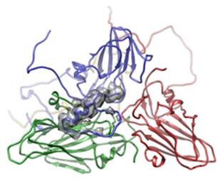 Fig. 1 Atomic model for SAT2 wildtype.1
Key structural properties of SAT2:
- Typical metal phosphodiesterase folding
- Double zinc ion catalytic center embedded hydrophobic pocket
- N-terminal SAP domain mediates membrane binding function
Functions of SAT2
The core function of the acidic sphingomyelinase encoded by the SAT2 gene is to catalyze the hydrolysis of sphingomyelin. However, this enzyme and its metabolites are also deeply involved in the regulation of various pathophysiological processes.
| Function | Description |
| Sphingomyelin hydrolysis | In the acidic environment of lysosomes, it specifically catalyzes sphingomipin to generate ceramides and phosphocholine, maintaining the lipid homeostasis of cell membranes. |
| Ceramide signal conduction | Ceramides produced by hydrolysis act as second messengers, regulating important signaling pathways such as apoptosis, autophagy and stress response. |
| Associated with atherosclerosis | Abnormal accumulation of sphingomyelin in vascular endothelial cells can promote lipid plaque formation, and the activity of this enzyme is positively correlated with the degree of atherosclerotic lesions. |
| The pathological basis of Niemann-Pick disease | The loss of enzyme activity leads to the inability of sphingomyelin to be degraded in mononuclear macrophage system organs, resulting in typical lipid accumulation symptoms. |
| Regulation of tumor drug resistance | Under the pressure of chemotherapy, the ceramide signaling axis mediated by this enzyme has been confirmed to affect the formation of drug resistance phenotypes in various cancer cells. |
The catalytic efficiency of this enzyme reaches its peak at pH 4.5-5.0. Its reaction kinetics conforms to the characteristics of the Michelin equation, but is significantly regulated by the membrane lipid microenvironment. This substrate interface activation mechanism is the key feature that distinguishes it from neutral sphingomipin enzymes.
Applications of SAT2 and SAT2 Antibody in Literature
1. Li, Qian, et al. "B and T cell epitopes of the incursionary foot-and-mouth disease virus serotype SAT2 for vaccine development." Viruses 15.3 (2023): 797. https://doi.org/10.3390/v15030797
The article indicates that foot-and-mouth disease SAT2 poses a threat to prevention and control due to the lack of cross-protection. Multi-epitope vaccines are an effective strategy to solve this problem, and their development relies on the bioinformatics identification of B and T cell epitopes. This article reviews the research progress of SAT2 epitopes and the experimental achievements of vaccines.
2. Mamabolo, Mpho Victoria, et al. "Production of foot-and-mouth disease virus SAT2 VP1 protein." AMB Express 10.1 (2020): 2. https://doi.org/10.1186/s13568-019-0938-7
The study successfully and efficiently expressed the SAT2 VP1 protein of foot-and-mouth disease in Escherichia coli, with a yield of 2.15 g/L. This recombinant protein can have a specific reaction with SAT2 antiserum, providing a new approach for differentiating SAT serotypes and replacing viral reagents.
3. Rathogwa, Ntungufhadzeni M., et al. "Efficacy of SAT2 foot-and-mouth disease vaccines formulated with Montanide ISA 206B and Quil-A Saponin adjuvants." Vaccines 9.9 (2021): 996. https://doi.org/10.3390/vaccines9090996
This study compared the compatibility effects of two adjuvants with heat-stable SAT2 vaccine antigens. The ISA 206B adjuvant vaccine can stimulate a stronger neutralizing antibody and cellular immune response, and remains effective against viruses that simulate antigenic drift, providing support for the development of long-acting multivalent vaccines.
4. Zhao, Ben, et al. "The Tumor-Suppressive Role of SAT2 in Pancreatic Cancer: Involvement in PI3K/Akt-MAPK Pathways and Immune Modulation." Current Issues in Molecular Biology 47.10 (2025): 872. https://doi.org/10.3390/cimb47100872
Research has found that the expression of SAT2 is significantly reduced in pancreatic cancer and is associated with prognosis. In vitro and in vivo experiments have confirmed that high expression of SAT2 can inhibit tumor proliferation and migration through pathways such as PI3K/Akt, and is associated with tumor immune infiltration, providing a new target for treatment.
5. Obinata, Hiroyuki, Asako Sugimoto, and Shinsuke Niwa. "Streptothricin acetyl transferase 2 (Sat2): A dominant selection marker for Caenorhabditis elegans genome editing." PLoS One 13.5 (2018): e0197128. https://doi.org/10.1371/journal.pone.0197128
This study established a novel antibiotic screening marker for nematodes based on the NTC and Sat2 genes. This system can efficiently screen transgenic individuals and can be combined with other systems, providing a powerful new tool for genetic manipulation.
Creative Biolabs: SAT2 Antibodies for Research
Creative Biolabs specializes in the production of high-quality SAT2 antibodies for research and industrial applications. Our portfolio includes monoclonal antibodies tailored for ELISA, Flow Cytometry, Western blot, immunohistochemistry, and other diagnostic methodologies.
- Custom SAT2 Antibody Development: Tailor-made solutions to meet specific research requirements.
- Bulk Production: Large-scale antibody manufacturing for industry partners.
- Technical Support: Expert consultation for protocol optimization and troubleshooting.
- Aliquoting Services: Conveniently sized aliquots for long-term storage and consistent experimental outcomes.
For more details on our SAT2 antibodies, custom preparations, or technical support, contact us at email.
Reference
- Kotecha, Abhay, et al. "Chimeric O1K foot-and-mouth disease virus with SAT2 outer capsid as an FMD vaccine candidate." Scientific Reports 8.1 (2018): 13654. https://doi.org/10.1038/s41598-018-31856-x
Anti-SAT2 antibodies
 Loading...


Hot products 
-
Mouse Anti-ADGRL2 Recombinant Antibody (V2-58519) (CBMAB-L0166-YJ)

-
Mouse Anti-ALDOA Recombinant Antibody (A2) (CBMAB-A2316-YC)

-
Mouse Anti-CCDC6 Recombinant Antibody (CBXC-0106) (CBMAB-C5397-CQ)

-
Mouse Anti-AQP2 Recombinant Antibody (E-2) (CBMAB-A3358-YC)

-
Mouse Anti-BZLF1 Recombinant Antibody (BZ.1) (CBMAB-AP705LY)

-
Mouse Anti-FPR2 Recombinant Antibody (1D6) (CBMAB-F2628-CQ)

-
Mouse Anti-APOE Recombinant Antibody (A1) (CBMAB-0078CQ)

-
Mouse Anti-APOA1 Monoclonal Antibody (CBFYR0637) (CBMAB-R0637-FY)

-
Mouse Anti-COL12A1 Recombinant Antibody (CBYY-C3117) (CBMAB-C4560-YY)

-
Mouse Anti-CAT Recombinant Antibody (724810) (CBMAB-C8431-LY)

-
Mouse Anti-FLI1 Recombinant Antibody (CBXF-0733) (CBMAB-F0435-CQ)

-
Mouse Anti-ENO2 Recombinant Antibody (H14) (CBMAB-E1341-FY)

-
Rabbit Anti-ALOX5AP Recombinant Antibody (CBXF-1219) (CBMAB-F0750-CQ)

-
Mouse Anti-ATG5 Recombinant Antibody (9H197) (CBMAB-A3945-YC)

-
Mouse Anti-FN1 Monoclonal Antibody (71) (CBMAB-1241CQ)

-
Mouse Anti-EIF4G1 Recombinant Antibody (2A9) (CBMAB-A2544-LY)

-
Mouse Anti-BAX Recombinant Antibody (CBYY-0216) (CBMAB-0217-YY)

-
Mouse Anti-ARIH1 Recombinant Antibody (C-7) (CBMAB-A3563-YC)

-
Mouse Anti-ACLY Recombinant Antibody (V2-179314) (CBMAB-A0610-YC)

-
Mouse Anti-DES Monoclonal Antibody (440) (CBMAB-AP1857LY)

- AActivation
- AGAgonist
- APApoptosis
- BBlocking
- BABioassay
- BIBioimaging
- CImmunohistochemistry-Frozen Sections
- CIChromatin Immunoprecipitation
- CTCytotoxicity
- CSCostimulation
- DDepletion
- DBDot Blot
- EELISA
- ECELISA(Cap)
- EDELISA(Det)
- ESELISpot
- EMElectron Microscopy
- FFlow Cytometry
- FNFunction Assay
- GSGel Supershift
- IInhibition
- IAEnzyme Immunoassay
- ICImmunocytochemistry
- IDImmunodiffusion
- IEImmunoelectrophoresis
- IFImmunofluorescence
- IGImmunochromatography
- IHImmunohistochemistry
- IMImmunomicroscopy
- IOImmunoassay
- IPImmunoprecipitation
- ISIntracellular Staining for Flow Cytometry
- LALuminex Assay
- LFLateral Flow Immunoassay
- MMicroarray
- MCMass Cytometry/CyTOF
- MDMeDIP
- MSElectrophoretic Mobility Shift Assay
- NNeutralization
- PImmunohistologyp-Paraffin Sections
- PAPeptide Array
- PEPeptide ELISA
- PLProximity Ligation Assay
- RRadioimmunoassay
- SStimulation
- SESandwich ELISA
- SHIn situ hybridization
- TCTissue Culture
- WBWestern Blot
