
SSR1 Antibodies
Background
The SSR1 gene encodes an endoplasmic reticulum membrane-integrated protein, which mainly participates in the post-translational transport of proteins and the endoplasmic reticulum-related degradation pathway. This protein acts as the subunit of the signal sequence receptor and plays a crucial role in the recognition process as the newly synthesized polypeptide chain enters the endoplasmic reticulum cavity. At the same time, it maintains the cellular protein homeostasis by regulating the reverse transport of misfolded proteins. Scientists first identified this gene through yeast genetic screening in the 1990s, and its functional research provided an important paradigm for the quality control mechanism of the secretion pathway. The highly conserved structure and function of this gene have become a classic model for studying endoplasmic reticulum stress, unfolded protein response, and related diseases, and have fundamental significance for understanding inter-organelle communication and the protein metabolic network.
Structure of SSR1
SSR1 is a signal recognition particle receptor protein with a molecular weight of approximately 20.5 kDa. This value may vary slightly among different species due to the slight adjustment of amino acid sequences.
| Species | Human | Mouse | Zebrafish | Arabidopsis thaliana |
| Molecular Weight (kDa) | 20.5 | 20.4 | 20.7 | 22.1 |
| Primary Structural Differences | The endoplasmic reticulum positioning signal is clear | Highly conserved in the transmembrane region | Similar core functional domains | There exist plant-specific extended sequences |
The protein encoded by this gene is composed of approximately 180 amino acids and exhibits a typical multi-pass transmembrane structure. Its primary structure contains several hydrophobic α-helical transmembrane regions, forming the core of the channel that penetrates the endoplasmic reticulum membrane. The key functional domains include the N-terminal domain facing the cytoplasm, which is responsible for interacting with the signal recognition particle (SRP); and the short tail at the C-terminus, which participates in signal transduction within the endoplasmic reticulum lumen. These structures work together to ensure that newly synthesized peptide chains can precisely target and translocate into the endoplasmic reticulum.
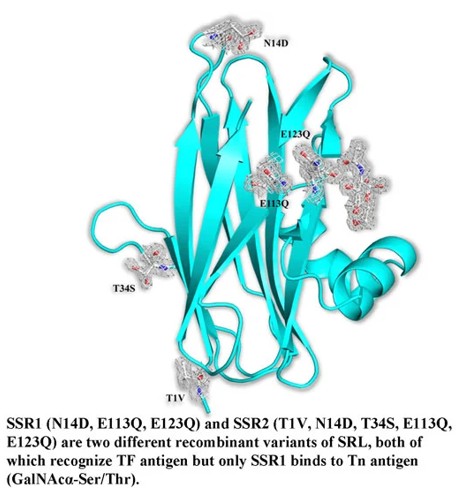 Fig. 1 Comparative Schematic of Mutations in SSR1 and SSR2 SRL Variants.1
Key structural properties of SSR1:
- Multiple transmembrane hydrophobic α-helix bundles structure
- Transmembrane helical form hydrophobic core, forms the endoplasm retinal integration channels
- The N-terminal domain facing the cytoplasm is responsible for identifying signal recognition particles
- Conservative acid residues involved in maintaining cluster channel charge balance and regulate transfer efficiency
Functions of SSR1
The core function of the SSR1 protein is to act as a signal recognition particle receptor, responsible for directing the ribosome‑nascent peptide chain complex to the endoplasmic reticulum membrane. At the same time, it also participates in regulating protein quality control and endoplasmic reticulum stress responses.
| Function | Description |
| Protein Transport | Identify and bind to the signal recognition particle-rubiscope complex in the cytoplasm, guiding newly synthesized peptide chains into the endoplasmic reticulum cavity for processing. |
| Channel Regulation by Translocation | Works in synergy with the Sec61 translocation complex to form and regulate the protein channel through which peptide chains pass through the endoplasmic reticulum membrane. |
| Translation Pause Adjustment | Its combination with SRP helps to temporarily slow down the translation speed, ensuring the coordination between translocation and translation. |
| Maintenance of Endoplasmic Reticulum Homeostasis | By ensuring the correct localization of secretory proteins and membrane proteins, it indirectly participates in maintaining the protein folding environment and homeostasis of the endoplasmic reticulum. |
| Disease Association | Its dysfunction is associated with certain genetic disorders and cancers, and may affect the cell's secretion capabilities and stress response. |
Unlike simple receptors that bind to a single substrate, the function of SSR1 relies on its precise collaboration with multiple dynamic complexes, demonstrating its pivotal role in the fundamental cellular process of co-translational transport.
Applications of SSR1 and SSR1 Antibody in Literature
1. Peppa, Vassiliki I., et al. "Molecular cloning, carbohydrate specificity and the crystal structure of two Sclerotium rolfsii lectin variants." Molecules 20.6 (2015): 10848-10865. https://doi.org/10.3390/molecules200610848
The article indicates that SRL is a lectin derived from the uniform small nucleus fungus, which can specifically recognize the TF antigen on the surface of cancer cells and inhibit tumors. The study constructed two recombinant variants of SRL, namely SSR1 and SSR2. Both of them retain the ability to bind to the TF antigen, but only SSR1 can bind to the Tn antigen. The crystal structure analysis revealed the fine sugar-binding specificity of it.
2. Zhang, Wen, et al. "Blood SSR1: A possible biomarker for early prediction of Parkinson’s disease." Frontiers in Molecular Neuroscience 15 (2022): 762544. https://doi.org/10.3389/fnmol.2022.762544
The research has found that the peripheral blood gene SSR1 can serve as a potential biomarker for the early diagnosis of Parkinson's disease. Its expression level is negatively correlated with the survival of dopaminergic neurons, and it has already increased before the onset of abnormal animal behaviors. Combined with an artificial intelligence model, it demonstrates excellent predictive value for the disease.
3. Li, Jing, et al. "PBX2-mediated circTLK1 activates JAK/STAT signaling to promote gliomagenesis via miR-452-5p/SSR1 axis." Frontiers in Genetics 12 (2021): 698831. https://doi.org/10.3389/fgene.2021.698831
This study found that circular RNA circTLK1 is highly expressed in glioma. It is regulated by the transcription factor PBX2 and can bind to miR-452-5p to upregulate SSR1, activate the JAK/STAT signaling pathway, and ultimately promote the progression of glioma.
4. Feng, Xuanjun, et al. "Plant-specific cochaperone SSR1 affects root elongation by modulating the mitochondrial iron-sulfur cluster assembly machinery." PLoS genetics 21.2 (2025): e1011597. https://doi.org/10.1371/journal.pgen.1011597
The research has found that the mitochondrial protein SSR1 in Arabidopsis thaliana, as a plant-specific factor, participates in the assembly of mitochondrial iron-sulfur clusters by cooperating with HSCA2 and ISU1. The absence of SSR1 leads to increased iron accumulation at the root tip and a decrease in the activity of iron-sulfur proteins, which affects root development. Its role becomes particularly important under environmental stress conditions.
5. Byrne, Alexander, et al. "Investigating in vivo Mycobacterium avium subsp. paratuberculosis microevolution and mixed strain infections." Microbiology Spectrum 11.5 (2023): e01716-23. https://doi.org/10.1128/spectrum.01716-23
The research found that variations in the short sequence repeat region of SSR1 in Mycobacterium paratuberculosis can alter the reading frame of the ORF1 gene, thereby affecting the structure of its surface proteins. This gene regulation, which is conserved within the Mycobacterium complex but occurs as a frameshift only in this specific bacterium, may affect its infectivity.
Creative Biolabs: SSR1 Antibodies for Research
Creative Biolabs specializes in the production of high-quality SSR1 antibodies for research and industrial applications. Our portfolio includes monoclonal antibodies tailored for ELISA, Flow Cytometry, Western blot, immunohistochemistry, and other diagnostic methodologies.
- Custom SSR1 Antibody Development: Tailor-made solutions to meet specific research requirements.
- Bulk Production: Large-scale antibody manufacturing for industry partners.
- Technical Support: Expert consultation for protocol optimization and troubleshooting.
- Aliquoting Services: Conveniently sized aliquots for long-term storage and consistent experimental outcomes.
For more details on our SSR1 antibodies, custom preparations, or technical support, contact us at email.
Reference
- Peppa, Vassiliki I., et al. "Molecular cloning, carbohydrate specificity and the crystal structure of two Sclerotium rolfsii lectin variants." Molecules 20.6 (2015): 10848-10865. Distributed under the same Creative Commons license CC BY 4.0 as the original. Cropped from the original figure. https://doi.org/10.3390/molecules200610848
Anti-SSR1 antibodies
 Loading...
 Compare




Hot products 
- AActivation
- AGAgonist
- APApoptosis
- BBlocking
- BABioassay
- BIBioimaging
- CImmunohistochemistry-Frozen Sections
- CIChromatin Immunoprecipitation
- CTCytotoxicity
- CSCostimulation
- DDepletion
- DBDot Blot
- EELISA
- ECELISA(Cap)
- EDELISA(Det)
- ESELISpot
- EMElectron Microscopy
- FFlow Cytometry
- FNFunction Assay
- GSGel Supershift
- IInhibition
- IAEnzyme Immunoassay
- ICImmunocytochemistry
- IDImmunodiffusion
- IEImmunoelectrophoresis
- IFImmunofluorescence
- IGImmunochromatography
- IHImmunohistochemistry
- IMImmunomicroscopy
- IOImmunoassay
- IPImmunoprecipitation
- ISIntracellular Staining for Flow Cytometry
- LALuminex Assay
- LFLateral Flow Immunoassay
- MMicroarray
- MCMass Cytometry/CyTOF
- MDMeDIP
- MSElectrophoretic Mobility Shift Assay
- NNeutralization
- PImmunohistologyp-Paraffin Sections
- PAPeptide Array
- PEPeptide ELISA
- PLProximity Ligation Assay
- RRadioimmunoassay
- SStimulation
- SESandwich ELISA
- SHIn situ hybridization
- TCTissue Culture
- WBWestern Blot
