CCL3 Antibodies
Background
CCL3 is a small molecule chemokine protein secreted by lymphocytes and macrophages. This gene mediates the chemotactic movement and activation of immune cells by binding to its receptors CCR1/CCR5, playing a core role in inflammatory responses and anti-pathogen infections. Research has found that CCL3 plays a key role in the suppression of HIV virus because it can competitively block the binding of the virus to the host cell receptor. This gene was first identified in 1988. The analysis of its tertiary structure provides an important foundation for the development of anti-inflammatory drugs targeting chemokine pathways and has a continuous promoting effect on the research of immune regulatory mechanisms and the development of disease treatment strategies.
Structure of CCL3
CCL3 is a relatively small protein with a molecular weight of approximately 8-10 kDa. This molecular weight may vary slightly among different species, mainly due to subtle changes in amino acid sequences.
| Species | Human | Mouse | Rat |
| Molecular Weight (kDa) | 8.0 | 8.7 | 8.9 |
| Primary Structural Differences | Conserved sequence, highly similar to most mammals | There are a few amino acid variations | Highly homologous to rat and mouse sequences |
The CCL3 protein is typically composed of about 70 to 92 amino acids and forms a typical chemokine folding structure. Its primary structure contains a conserved tetracysteine motif (CCL motif), which stabilizes the overall conformation through disulfide bonds. The core functional region of this protein can specifically bind to G protein-coupled receptors (such as CCR1 and CCR5), thereby mediating the chemotactic activities of immune cells. Its tertiary structure is mainly composed of anti-parallel β -folding and a C-terminal α -helix, forming a hydrophobic binding pocket, which is crucial for its recognition with the receptor and signal transduction.
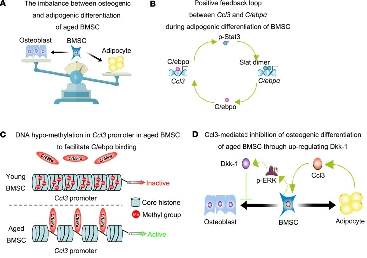 Fig. 1 Potential role of CCL3 in bone loss and bone marrow adiposity of aged mice.1
Key structural properties of CCL3:
- Conservative chemokine folding structure
- Core through the disulfide bond (such as Cys10 - Cys34) three-dimensional structure stability
- The N-terminal domain with specific recognition of CCR1/CCR5 receptors
Functions of CCL3
The main function of CCL3 is to mediate the chemotaxis and activation of immune cells. In addition, it is widely involved in key physiological and pathological processes such as inflammatory responses, host anti-infection and immune regulation.
| Function | Description |
| Chemotaxis of immune cells | Recruit monocytes, macrophages, neutrophils and T cells to the site of infection or inflammation to coordinate the early immune response. |
| Activation of immune cells | By binding to the receptors CCR1/CCR5, it activates signaling pathways such as MAPK and PI3K, promoting the proliferation, differentiation of immune cells and the release of cytokines. |
| Antiviral response | In HIV infection, it can competitively bind to the CCR5 receptor, inhibit the entry of the virus into host cells, and has a certain viral inhibitory function. |
| Inflammatory regulation | Participate in the occurrence and development of various acute and chronic inflammatory diseases (such as rheumatoid arthritis and asthma), and promote or inhibit the inflammatory process. |
| Tissue repair and regeneration | In the later stage of injury and infection, it assists in eliminating pathogens, regulates fibroblast activation and angiogenesis, and affects tissue remodeling and repair. |
The dose-effect curve of CCL3 binding to its receptor usually shows typical saturation characteristics, indicating that it has highly efficient chemotactic activity at low concentrations and may trigger desensitization reactions at high concentrations, thereby precisely regulating the migration process of immune cells.
Applications of CCL3 and CCL3 Antibody in Literature
1. Wei, Yan-Fei, et al. "Helicobacter pylori disrupts gastric mucosal homeostasis by stimulating macrophages to secrete CCL3." Cell Communication and Signaling 22.1 (2024): 263. https://doi.org/10.1186/s12964-024-01627-5
The article indicates that Helicobacter pylori infection promotes the secretion of CCL3 by macrophages through the JAK1-STAT1 pathway, and then disrupts the tight junctions of gastric epithelial cells by activating P38 phosphorylation, leading to gastric mucosal damage.
2. Li, Hanwen, et al. "Lumbar instability remodels cartilage endplate to induce intervertebral disc degeneration by recruiting osteoclasts via Hippo-CCL3 signaling." Bone Research 12.1 (2024): 34. https://doi.org/10.1038/s41413-024-00331-x
The article indicates that lumbar instability activates the expression of CCL3 in the cartilage endplate through the Hippo signaling pathway, which in turn recruit osteoclasts to cause cheid-like changes in the endplate and intervertebral disc degeneration. Overexpression of Yap1 can inhibit the transcription of CCL3 and reverse this process.
3. Ishida, Yuko, et al. "Prevention of CaCl2-induced aortic inflammation and subsequent aneurysm formation by the CCL3–CCR5 axis." Nature communications 11.1 (2020): 5994. https://doi.org/10.1038/s41467-020-19763-0
The article indicates that CCL3 inhibits macrophage infiltration and MMP-9 expression by binding to the receptor CCR5, thereby suppressing the formation of abdominal aortic aneurysms induced by calcium chloride or angiotensin II. Exogenous CCL3 treatment can delay the progression of aneurysms.
4. Ma, aoqiang, et al. "CCL3 Promotes Proliferation of Colorectal Cancer Related with TRAF6/NF‐κB Molecular Pathway." Contrast media & molecular imaging 2022.1 (2022): 2387192. https://doi.org/10.1155/2022/2387192
The article indicates that CCL3 is highly expressed in colorectal cancer, promoting tumor proliferation and invasion by activating the TRAF6/NF-κB pathway. Its expression is related to clinical stage and nerve invasion, and is positively correlated with the receptor CCR5.
5. Yu, Degang, et al. "CCL3 in the bone marrow microenvironment causes bone loss and bone marrow adiposity in aged mice." JCI insight 8.1 (2023): e159107. https://doi.org/10.1172/jci.insight.159107
The article indicates that CCL3 accumulates in the serum of aging mice, promotes adipogenic differentiation of bone marrow stromal cells through the STAT3/C/EBPα positive feedback loop, and inhibits osteogenic differentiation by suppressing β-catenin activity through ERK/DKK1. Blocking CCL3 can improve the bone aging phenotype.
Creative Biolabs: CCL3 Antibodies for Research
Creative Biolabs specializes in the production of high-quality CCL3 antibodies for research and industrial applications. Our portfolio includes monoclonal antibodies tailored for ELISA, Flow Cytometry, Western blot, immunohistochemistry, and other diagnostic methodologies.
- Custom CCL3 Antibody Development: Tailor-made solutions to meet specific research requirements.
- Bulk Production: Large-scale antibody manufacturing for industry partners.
- Technical Support: Expert consultation for protocol optimization and troubleshooting.
- Aliquoting Services: Conveniently sized aliquots for long-term storage and consistent experimental outcomes.
For more details on our CCL3 antibodies, custom preparations, or technical support, contact us at email.
Reference
- Yu, Degang, et al. "CCL3 in the bone marrow microenvironment causes bone loss and bone marrow adiposity in aged mice." JCI insight 8.1 (2023): e159107. https://doi.org/10.1172/jci.insight.159107
Anti-CCL3 antibodies
 Loading...


Hot products 
-
Mouse Anti-8-oxoguanine Recombinant Antibody (V2-7697) (CBMAB-1869CQ)

-
Mouse Anti-ARSA Recombinant Antibody (CBYC-A799) (CBMAB-A3679-YC)

-
Mouse Anti-Acetyl-α-Tubulin (Lys40) Recombinant Antibody (V2-623485) (CBMAB-CP2897-LY)

-
Mouse Anti-CD33 Recombinant Antibody (P67.6) (CBMAB-C10189-LY)

-
Mouse Anti-CD24 Recombinant Antibody (SN3) (CBMAB-C1037-CQ)

-
Mouse Anti-AGO2 Recombinant Antibody (V2-634169) (CBMAB-AP203LY)

-
Mouse Anti-CCNH Recombinant Antibody (CBFYC-1054) (CBMAB-C1111-FY)

-
Rabbit Anti-CCL5 Recombinant Antibody (R0437) (CBMAB-R0437-CN)

-
Mouse Anti-CRTAM Recombinant Antibody (CBFYC-2235) (CBMAB-C2305-FY)

-
Mouse Anti-FOSB Recombinant Antibody (CBXF-3593) (CBMAB-F2522-CQ)

-
Rat Anti-CD300A Recombinant Antibody (172224) (CBMAB-C0423-LY)

-
Mouse Anti-DDC Recombinant Antibody (8E8) (CBMAB-0992-YC)

-
Mouse Anti-FLT1 Recombinant Antibody (11) (CBMAB-V0154-LY)

-
Mouse Anti-CD24 Recombinant Antibody (ALB9) (CBMAB-0176CQ)

-
Mouse Anti-C5b-9 Recombinant Antibody (aE11) (CBMAB-AO138LY)

-
Mouse Anti-ARHGDIA Recombinant Antibody (CBCNA-009) (CBMAB-R0415-CN)

-
Mouse Anti-CCDC25 Recombinant Antibody (CBLC132-LY) (CBMAB-C9786-LY)

-
Mouse Anti-DHFR Recombinant Antibody (D0821) (CBMAB-D0821-YC)

-
Mouse Anti-ADAM12 Recombinant Antibody (V2-179752) (CBMAB-A1114-YC)

-
Mouse Anti-GFAP Recombinant Antibody (20) (CBMAB-G2914-LY)

- AActivation
- AGAgonist
- APApoptosis
- BBlocking
- BABioassay
- BIBioimaging
- CImmunohistochemistry-Frozen Sections
- CIChromatin Immunoprecipitation
- CTCytotoxicity
- CSCostimulation
- DDepletion
- DBDot Blot
- EELISA
- ECELISA(Cap)
- EDELISA(Det)
- ESELISpot
- EMElectron Microscopy
- FFlow Cytometry
- FNFunction Assay
- GSGel Supershift
- IInhibition
- IAEnzyme Immunoassay
- ICImmunocytochemistry
- IDImmunodiffusion
- IEImmunoelectrophoresis
- IFImmunofluorescence
- IGImmunochromatography
- IHImmunohistochemistry
- IMImmunomicroscopy
- IOImmunoassay
- IPImmunoprecipitation
- ISIntracellular Staining for Flow Cytometry
- LALuminex Assay
- LFLateral Flow Immunoassay
- MMicroarray
- MCMass Cytometry/CyTOF
- MDMeDIP
- MSElectrophoretic Mobility Shift Assay
- NNeutralization
- PImmunohistologyp-Paraffin Sections
- PAPeptide Array
- PEPeptide ELISA
- PLProximity Ligation Assay
- RRadioimmunoassay
- SStimulation
- SESandwich ELISA
- SHIn situ hybridization
- TCTissue Culture
- WBWestern Blot
