CCR2 Antibodies
Background
CCR2 is a G protein-coupled receptor, mainly expressed on the surface of immune cells such as monocytes and dendritic cells. The protein encoded by this gene mediates the migration of immune cells to the inflammatory site by specifically binding to chemokines (such as CCL2), playing a core role in immune response and inflammatory regulation. Research has found that CCR2 is closely related to a variety of diseases, including autoimmune diseases, atherosclerosis and cancer metastasis, etc. This receptor was first cloned in 1994. Its seven-transmembrane structure feature makes it an important target for drug development. Currently, several CCR2 antagonists have entered the clinical trial stage. In-depth research on the CCR2 signaling pathway not only promotes the development of therapeutic strategies for inflammatory diseases but also provides an important model for understanding the functional mechanisms of the GPCR family.
Structure of CCR2
CCR2 is a G protein-coupled receptor with a molecular weight of approximately 41-46 kDa, and its precise molecular weight varies slightly due to the degree of glycosylation and species differences.
| Species | Human | Mouse | Rat | Non-human primates |
| Molecular Weight (kDa) | 42.3 | 41.8 | 41.9 | 42.1 |
| Primary Structural Differences | Containing 368 amino acids, N side has multiple glycosylation sites | 76% homology with human | 75% homology with human | Highly conserved to the human sequence |
The CCR2 protein is composed of approximately 368 amino acids and features a typical seven-transmembrane structure. This receptor recognizes chemokine ligands (such as CCL2) through the N-terminal extracellular domain, and the third intracellular loop couples with the G protein to transmit signals. Its transmembrane region forms conserved ligand binding pockets, and the aspartic acid residues in the second and third transmembrane regions are crucial for ligand binding. The C-terminal contains multiple serine/threonine phosphorylation sites, which are involved in the regulation of receptor internalization and desensitization.
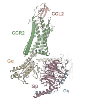 Fig. 1 Models of the CCL2–CCR2–Gi.1
Key structural properties of CCR2:
- Typical seventh-transmembrane domains (GPCR superfamily)
- Extracellular N-terminal region responsible for chemokines recognition and combination
- Conserved DRY motif is located in the second intracellular ring, mediating G protein signal transduction
Functions of CCR2
The main function of CCR2 is to mediate the migration of immune cells and inflammatory responses. In addition, it is also involved in a variety of pathophysiological processes, including autoimmune diseases, cancer metastasis and metabolic disorders.
| Function | Description |
| Chemotaxis of immune cells | CCR2 guides monocytes and dendritic cells to migrate to the inflammatory site by binding to chemokines such as CCL2. |
| Regulation of inflammatory response | When there is infection or tissue damage, CCR2 activates downstream signaling pathways, promoting the release of inflammatory factors and the initiation of immune responses. |
| Participation in disease development | Participate in the occurrence of diseases such as atherosclerosis, rheumatoid arthritis, promote immune cells and tissue damage. |
| Tumor microenvironment formation | Assist in the recruitment of tumor-associated macrophages (Tams) in tumor models, and promote tumor growth and metastasis. |
| Metabolic regulation | Effect of adipose tissue inflammation and insulin sensitivity, associated with metabolic diseases such as obesity and diabetes. |
The signal response of CCR2 has the typical characteristics of G protein-coupled receptors. Its ligand binding curve shows saturation kinetics characteristics, indicating that it can be effectively activated at low ligand concentrations and is suitable for sensitively regulating the directional movement of immune cells.
Applications of CCR2 and CCR2 Antibody in Literature
1. Tian, Qi, et al. "Inhibition of CCR2 attenuates neuroinflammation and neuronal apoptosis after subarachnoid hemorrhage through the PI3K/Akt pathway." Journal of neuroinflammation 19.1 (2022): 312. https://doi.org/10.1186/s12974-022-02676-8
This study, through bioinformatics analysis and experimental verification, found that the expression of CCR2 is upregulated after subarachnoid hemorrhage (SAH). Inhibiting CCR2 can alleviate neuroinflammation and neuronal apoptosis through the PI3K/AKT pathway and improve prognosis. The level of CCR2 in cerebrospinal fluid has predictive value for the 6-month outcome of patients.
2. Wang, Xinyue, et al. "Rescue RM/CS-AKI by blocking strategy with one-dose anti-myoglobin RabMAb." Nature Communications 16.1 (2025): 1044. https://doi.org/10.1007/s43440-021-00280-w
The article indicates that the CCL2-CCR2 axis not only participates in neuroinflammation but also influences emotional disorders by regulating homeostatic functions such as neurogenesis and transmission. Its functional disorder rather than simple inflammation is the underlying mechanism of depression and has become a new target for antidepressant treatment.
3. Fei, Liyang, et al. "Targeting the CCL2/CCR2 axis in cancer immunotherapy: one stone, three birds?." Frontiers in immunology 12 (2021): 771210. https://doi.org/10.3389/fimmu.2021.771210
The article indicates that CCR2 antagonists enhance the anti-tumor immune response by inhibiting the tumor infiltration of immunosuppressive monocytes/macrophages and Treg cells, and directly suppressing the survival and metastasis of tumor cells.
4. Kadomoto, Suguru, Kouji Izumi, and Atsushi Mizokami. "Roles of CCL2-CCR2 axis in the tumor microenvironment." International Journal of Molecular Sciences 22.16 (2021): 8530. https://doi.org/10.3390/ijms22168530
The article indicates that the CCL2-CCR2 signaling axis influences cancer progression by promoting tumor proliferation, invasion, angiogenesis, and recruiting immunosuppressive cells to shape the microenvironment, making it a potential therapeutic target.
5. Saadane, Aicha, et al. "CCR2-positive monocytes contribute to the pathogenesis of early diabetic retinopathy in mice." Diabetologia 66.3 (2023): 590-602. https://doi.org/10.1007/s00125-022-05860-w
The article indicates that CCR2+ inflammatory monocytes exacerbate capillary degeneration in diabetic retinopathy by promoting oxidative stress, inflammation and cytotoxicity. The absence of CCR2 can significantly inhibit this pathological process.
Creative Biolabs: CCR2 Antibodies for Research
Creative Biolabs specializes in the production of high-quality CCR2 antibodies for research and industrial applications. Our portfolio includes monoclonal antibodies tailored for ELISA, Flow Cytometry, Western blot, immunohistochemistry, and other diagnostic methodologies.
- Custom CCR2 Antibody Development: Tailor-made solutions to meet specific research requirements.
- Bulk Production: Large-scale antibody manufacturing for industry partners.
- Technical Support: Expert consultation for protocol optimization and troubleshooting.
- Aliquoting Services: Conveniently sized aliquots for long-term storage and consistent experimental outcomes.
For more details on our CCR2 antibodies, custom preparations, or technical support, contact us at email.
Reference
- Shao, Zhehua, et al. "Molecular insights into ligand recognition and activation of chemokine receptors CCR2 and CCR3." Cell Discovery 8.1 (2022): 44. https://doi.org/10.1038/s41421-022-00403-4
Anti-CCR2 antibodies
 Loading...


Hot products 
-
Rabbit Anti-ABL1 (Phosphorylated Y185) Recombinant Antibody (V2-443434) (PTM-CBMAB-0001YC)

-
Mouse Anti-ADGRE5 Recombinant Antibody (V2-360335) (CBMAB-C2088-CQ)

-
Mouse Anti-BZLF1 Recombinant Antibody (BZ.1) (CBMAB-AP705LY)

-
Mouse Anti-ARHGAP5 Recombinant Antibody (54/P190-B) (CBMAB-P0070-YC)

-
Mouse Anti-DLL4 Recombinant Antibody (D1090) (CBMAB-D1090-YC)

-
Mouse Anti-ADRB2 Recombinant Antibody (V2-180026) (CBMAB-A1420-YC)

-
Mouse Anti-ABL2 Recombinant Antibody (V2-179121) (CBMAB-A0364-YC)

-
Mouse Anti-BCL6 Recombinant Antibody (CBYY-0435) (CBMAB-0437-YY)

-
Rabbit Anti-AKT2 (Phosphorylated S474) Recombinant Antibody (V2-556130) (PTM-CBMAB-0605LY)

-
Mouse Anti-BRD3 Recombinant Antibody (CBYY-0801) (CBMAB-0804-YY)

-
Rabbit Anti-AKT3 Recombinant Antibody (V2-12567) (CBMAB-1057-CN)

-
Mouse Anti-ADIPOR1 Recombinant Antibody (V2-179982) (CBMAB-A1368-YC)

-
Mouse Anti-CD63 Recombinant Antibody (CBXC-1200) (CBMAB-C1467-CQ)

-
Mouse Anti-FOXA3 Recombinant Antibody (2A9) (CBMAB-0377-YC)

-
Rat Anti-AChR Recombinant Antibody (V2-12500) (CBMAB-0990-CN)

-
Mouse Anti-AQP2 Recombinant Antibody (G-3) (CBMAB-A3359-YC)

-
Rat Anti-ADGRE4 Recombinant Antibody (V2-160163) (CBMAB-F0011-CQ)

-
Rat Anti-ABCC11 Recombinant Antibody (V2-179001) (CBMAB-A0236-YC)

-
Mouse Anti-8-oxoguanine Recombinant Antibody (V2-7697) (CBMAB-1869CQ)

-
Mouse Anti-ENO1 Recombinant Antibody (CBYC-A950) (CBMAB-A4388-YC)

- AActivation
- AGAgonist
- APApoptosis
- BBlocking
- BABioassay
- BIBioimaging
- CImmunohistochemistry-Frozen Sections
- CIChromatin Immunoprecipitation
- CTCytotoxicity
- CSCostimulation
- DDepletion
- DBDot Blot
- EELISA
- ECELISA(Cap)
- EDELISA(Det)
- ESELISpot
- EMElectron Microscopy
- FFlow Cytometry
- FNFunction Assay
- GSGel Supershift
- IInhibition
- IAEnzyme Immunoassay
- ICImmunocytochemistry
- IDImmunodiffusion
- IEImmunoelectrophoresis
- IFImmunofluorescence
- IGImmunochromatography
- IHImmunohistochemistry
- IMImmunomicroscopy
- IOImmunoassay
- IPImmunoprecipitation
- ISIntracellular Staining for Flow Cytometry
- LALuminex Assay
- LFLateral Flow Immunoassay
- MMicroarray
- MCMass Cytometry/CyTOF
- MDMeDIP
- MSElectrophoretic Mobility Shift Assay
- NNeutralization
- PImmunohistologyp-Paraffin Sections
- PAPeptide Array
- PEPeptide ELISA
- PLProximity Ligation Assay
- RRadioimmunoassay
- SStimulation
- SESandwich ELISA
- SHIn situ hybridization
- TCTissue Culture
- WBWestern Blot
