CLDN5 Antibodies
Background
CLDN5 is a small molecule transmembrane protein that is mainly found at the tight junctions of vascular endothelial cells in vertebrates. This protein regulates vascular permeability by forming intercellular barrier structures and maintains the selective permeability functions of the blood-brain barrier and blood-tissue barrier. The nervous system relies on CLDN5 to strictly control the entry and exit of molecules through cerebral microvessels, ensuring the stability of the neural microenvironment. This gene was first identified in 1998, and the protein encoded by it is one of the most intensively studied members of the closed protein family. Its four transmembrane domains and the classical topology structure formed by the extracellular loops have become the paradigm for the study of tight junction proteins. The regulatory mechanisms of barrier functions under physiological and pathological conditions have been extensively explored, providing a key molecular basis for understanding paracellular transport, barrier integrity, and related disease mechanisms.
Structure of CLDN5
CLDN5 is a relatively small protein with a molecular weight of approximately 23 kDa. This molecular weight may vary slightly among different species due to minor differences in amino acid sequences.
| Species | Human | Mouse | Rat | Bovine |
| Molecular Weight (kDa) | 23 | 22.8 | 23.1 | 23.2 |
| Primary Structural Differences | Standard structure, containing four transmembrane domains | Highly conserved in the transmembrane region | Minor variations exist in the region of the extracellular loop | Has an extremely high homology with human protein sequences |
This protein is encoded by the human CLDN5 gene and typically consists of approximately 218 amino acid residues. It folds through its primary structure to form a tight junction protein core scaffold consisting of four transmembrane domains. Its structure contains two extracellular loops: the first extracellular loop is a key functional domain that forms the intercellular tight junction barrier and determines ion selectivity; the second extracellular loop is also involved in intercellular interactions. The secondary structure of CLDN5 is mainly composed of α-helices, which are oriented in the cell membrane and together form a hydrophilic channel microenvironment to filter specific small molecule ions. The N-terminus and C-terminus are both located in the cytoplasm, and they are stabilized at the tight junction by binding to intracellular scaffold proteins (such as ZO-1) and conduct regulatory signals.
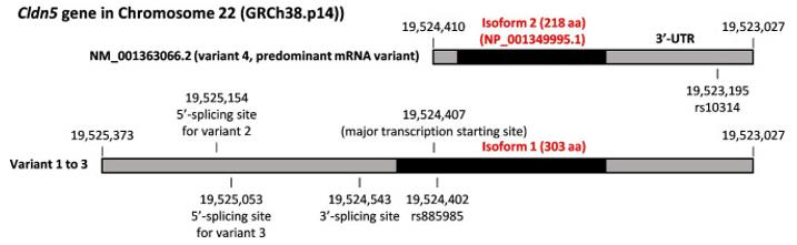 Fig. 1 The basic information of CLDN5 gene.1
Key structural properties of CLDN5:
- The core framework is composed of four transmembrane α-helices.
- Two extracellular rings, the first of which is a key area for barrier formation and ion screening
- The C-terminus contains a PDZ-binding domain for anchoring intracellular cytoskeletal proteins
- Charge selectivity through the charged amino acid residue on the first extracellular loop
Functions of CLDN5
The main function of the CLDN5 protein is to form a tight junction barrier between vascular endothelial cells and regulate vascular permeability. Additionally, it is involved in various pathological and physiological processes, including the regulation of immune cell migration and tumor metastasis.
| Function | Description |
| Barrier Formation | It forms a continuous physical barrier by interconnecting with the homologous CLDN5 proteins on adjacent endothelial cells, serving as the structural basis for biological barriers such as the blood-brain barrier. |
| Permeability Regulation | It selectively allows water, small ions, and specific solutes to pass through the cell bypass pathway, while strictly restricting the entry of large molecules and harmful substances into the tissue. |
| Maintenance of cell polarity | By connecting to the PDZ-binding domain at its cytoplasmic end with scaffold proteins (such as ZO-1), it helps establish and maintain the apical-basal lateral polarity of endothelial cells. |
| Signal Transduction Involvement | Its intracellular domain can serve as a platform to facilitate the integration of extracellular environmental signals with the rearrangement of the actin cytoskeleton within the cell. |
| Disease-related Effects | Its abnormal expression or function is closely related to processes such as blood-brain barrier disruption (e.g. stroke, brain tumor), vasculitic edema, and cancer metastasis. |
Unlike other tight junction proteins with complex regulation and multiple subtype combinations (such as Occludin), the channels formed by CLDN5 exhibit relative selectivity for small cations (such as Na⁺, K⁺), and their barrier function is direct and crucial, making them a core molecule for maintaining the stability of the microenvironment in the central nervous system.
Applications of CLDN5 and CLDN5 Antibody in Literature
1. Hashimoto, Yosuke, et al. "The CLDN5 gene at the blood-brain barrier in health and disease." Fluids and Barriers of the CNS 20.1 (2023): 22. https://doi.org/10.1186/s12987-023-00424-5
The article indicates that the CLDN5 gene encodes the tight junction protein CLDN5 of the blood-brain barrier. Its down-regulation leads to impaired barrier function and increases the risk of neurological and mental disorders, epilepsy, and dementia. The latest research has revealed its regulatory mechanism, the first case of functional gain-of-function mutation, and related treatment strategies.
2. Sun, Hui, et al. "Loss of CLDN5 in podocytes deregulates WIF1 to activate WNT signaling and contributes to kidney disease." Nature Communications 13.1 (2022): 1600. https://doi.org/10.1038/s41467-022-29277-6
The research has found that CLDN5 in podocytes stabilizes ZO1 and inhibits the nuclear entry of ZONAB, maintaining WIF1 expression to inhibit the WNT pathway. The absence of CLDN5 will exacerbate diabetic nephropathy and renal fibrosis, while exogenous supplementation of WIF1 can delay the progression of the disease.
3. Han, Lu, et al. "CLDN5 identified as a biomarker for metastasis and immune infiltration in gastric cancer via pan-cancer analysis." Aging (Albany NY) 15.11 (2023): 5032. https://doi.org/10.18632/aging.204776
The research has found that CLDN5 is abnormally expressed in various cancers and is associated with tumor stage, immune infiltration, and patient prognosis. The study has confirmed that it can serve as a potential diagnostic marker for gastric cancer and suggests that it may play an important role in the tumor immune microenvironment and immunotherapy.
4. Zhu, Xin-yu, et al. "CD34+ CLDN5+ tumor associated senescent endothelial cells through IGF2-IGF2R signaling increased cholangiocellular phenotype in hepatocellular carcinoma." Journal of Advanced Research (2024). https://doi.org/10.1016/j.jare.2024.12.008
The study found that in hepatocellular carcinoma, the senescent endothelial cell subpopulation (CD34+CLDN5+) recruits mesenchymal stem cells into the tumor microenvironment by secreting IGF2, promoting the characteristics of cancer stem cells and the formation of bile duct cell phenotypes, and exacerbating the malignant progression of the tumor.
5. Tesch, Florian, et al. "Super‐resolved local recruitment of CLDN5 to filtration slits implicates a direct relationship with podocyte foot process effacement." Journal of cellular and molecular medicine 25.16 (2021): 7631-7641. https://doi.org/10.1111/jcmm.16519
The study found that in various glomerular diseases, the area of podocyte damage would locally upregulate the tight junction protein CLDN5. The ratio of CLDN5 to nephrin was significantly correlated with the density of filtration gaps, suggesting that CLDN5 may serve as a biomarker for predicting early podocyte damage.
Creative Biolabs: CLDN5 Antibodies for Research
Creative Biolabs specializes in the production of high-quality CLDN5 antibodies for research and industrial applications. Our portfolio includes monoclonal antibodies tailored for ELISA, Flow Cytometry, Western blot, immunohistochemistry, and other diagnostic methodologies.
- Custom CLDN5 Antibody Development: Tailor-made solutions to meet specific research requirements.
- Bulk Production: Large-scale antibody manufacturing for industry partners.
- Technical Support: Expert consultation for protocol optimization and troubleshooting.
- Aliquoting Services: Conveniently sized aliquots for long-term storage and consistent experimental outcomes.
For more details on our CLDN5 antibodies, custom preparations, or technical support, contact us at email.
Reference
- Hashimoto, Yosuke, et al. "The CLDN5 gene at the blood-brain barrier in health and disease." Fluids and Barriers of the CNS 20.1 (2023): 22. Distributed under Open Access license CC BY 4.0, without modification.https://doi.org/10.1186/s12987-023-00424-5
Anti-CLDN5 antibodies
 Loading...


Hot products 
-
Mouse Anti-AAV-5 Recombinant Antibody (V2-503417) (CBMAB-V208-1369-FY)

-
Mouse Anti-APOE Recombinant Antibody (A1) (CBMAB-0078CQ)

-
Mouse Anti-APP Recombinant Antibody (DE2B4) (CBMAB-1122-CN)

-
Mouse Anti-DHFR Recombinant Antibody (D0821) (CBMAB-D0821-YC)

-
Mouse Anti-AMIGO2 Recombinant Antibody (CBYY-C0756) (CBMAB-C2192-YY)

-
Mouse Anti-GFAP Recombinant Antibody (20) (CBMAB-G2914-LY)

-
Rabbit Anti-DLK1 Recombinant Antibody (9D8) (CBMAB-D1061-YC)

-
Mouse Anti-ATP1B1 Recombinant Antibody (E4) (CBMAB-0463-LY)

-
Mouse Anti-AAV-5 Recombinant Antibody (V2-503416) (CBMAB-V208-1402-FY)

-
Mouse Anti-NSUN6 Recombinant Antibody (D-5) (CBMAB-N3674-WJ)

-
Rat Anti-4-1BB Recombinant Antibody (V2-1558) (CBMAB-0953-LY)

-
Mouse Anti-AFM Recombinant Antibody (V2-634159) (CBMAB-AP185LY)

-
Mouse Anti-BRD3 Recombinant Antibody (CBYY-0801) (CBMAB-0804-YY)

-
Mouse Anti-CECR2 Recombinant Antibody (CBWJC-2465) (CBMAB-C3533WJ)

-
Mouse Anti-FAS2 Monoclonal Antibody (1D4) (CBMAB-0071-CN)

-
Mouse Anti-DLL4 Recombinant Antibody (D1090) (CBMAB-D1090-YC)

-
Mouse Anti-C4B Recombinant Antibody (CBYY-C2996) (CBMAB-C4439-YY)

-
Mouse Anti-C5B-9 Recombinant Antibody (CBFYA-0216) (CBMAB-X0304-FY)

-
Mouse Anti-BSN Recombinant Antibody (219E1) (CBMAB-1228-CN)

-
Mouse Anti-DISP2 Monoclonal Antibody (F66A4B1) (CBMAB-1112CQ)

- AActivation
- AGAgonist
- APApoptosis
- BBlocking
- BABioassay
- BIBioimaging
- CImmunohistochemistry-Frozen Sections
- CIChromatin Immunoprecipitation
- CTCytotoxicity
- CSCostimulation
- DDepletion
- DBDot Blot
- EELISA
- ECELISA(Cap)
- EDELISA(Det)
- ESELISpot
- EMElectron Microscopy
- FFlow Cytometry
- FNFunction Assay
- GSGel Supershift
- IInhibition
- IAEnzyme Immunoassay
- ICImmunocytochemistry
- IDImmunodiffusion
- IEImmunoelectrophoresis
- IFImmunofluorescence
- IGImmunochromatography
- IHImmunohistochemistry
- IMImmunomicroscopy
- IOImmunoassay
- IPImmunoprecipitation
- ISIntracellular Staining for Flow Cytometry
- LALuminex Assay
- LFLateral Flow Immunoassay
- MMicroarray
- MCMass Cytometry/CyTOF
- MDMeDIP
- MSElectrophoretic Mobility Shift Assay
- NNeutralization
- PImmunohistologyp-Paraffin Sections
- PAPeptide Array
- PEPeptide ELISA
- PLProximity Ligation Assay
- RRadioimmunoassay
- SStimulation
- SESandwich ELISA
- SHIn situ hybridization
- TCTissue Culture
- WBWestern Blot
