DLC1 Antibodies
Background
The DLC1 gene encodes an Rho GTPase activator protein, which is mainly present in various tissue cells of mammals. This protein participates in key processes such as cytoskeletal recombination, cell migration and adhesion by regulating the activity of Rho family GTPases, and plays an important role in maintaining tissue homeostasis. In tumor research, DLC1 is widely regarded as an important tumor suppressor gene, and its expression loss is closely related to the metastasis of various cancers such as liver cancer and breast cancer. This gene was first identified in human liver cancer cells by a research team in 1998. The protein it encodes, due to its unique cellular regulatory mechanism, has become an important model for the study of cancer treatment targets, providing a key theoretical basis for understanding cell signal transduction and tumor suppression mechanisms.
Structure of DLC1
The molecular weight of the protein encoded by the DLC1 gene is approximately 120 kDa, and its specific size may vary depending on different splicing variants. This protein has highly conserved domains in different species, but amino acid sequence variations in specific regions may affect its interaction with signal proteins. The following is a comparison of the molecular characteristics of the DLC1 protein in some species.
| Species | Human | Mouse | Rat | Zebrafish |
| Molecular Weight (kDa) | 120 | 118 | 119 | 115 |
| Primary Structural Differences | Contains the SAM and RhoGAP domains | SAM structure domain highly conservative | Similar GAP activity area | Core structure domain, the carboxyl end shorter |
The DLC1 protein is composed of approximately 1090 amino acids, and its primary structure mainly consists of the SAM domain at the amino terminal, the RhoGAP catalytic domain in the center, and the Start-like domain at the carboxyl terminal. The typical structural feature of this protein is the formation of a highly conserved GAP catalytic domain in the central region. This domain maintains a three-dimensional conformation through a hydrophobic core and directly binds to the active site of the Rho family GTP enzyme. The GTPase activation function of DLC1 mainly depends on the catalytic pocket composed of arginine residues within its GAP domain. These residues interact with the switch II region of GTPase to accelerate the hydrolysis of GTP to GDP. The Start-like domain near the carboxyl terminus is responsible for sensing lipid signals and regulating the membrane localization of proteins, while the SAM domain is involved in protein dimerization and interactions with other signaling molecules, jointly fulfilling the regulatory function of cytoskeletal rearrangement.
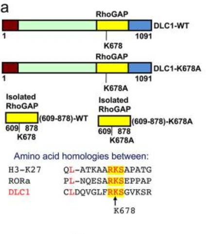 Fig. 1 Schematic representation of full-length DLC1 domains.1
Key structural properties of DLC1:
- Contains highly conserved SAM protein interaction domains
- The central structure domain RhoGAP catalytic activity center
- Carboxy-terminal start-like domains are involved in lipid binding
- Specific residues precisely control the activity of Rho GTP enzyme through allosteric regulation
Functions of DLC1
The main function of the protein encoded by the DLC1 gene is to act as a negative regulatory factor of Rho GTP enzyme, inhibiting cell proliferation and migration. At the same time, it is also involved in a variety of cellular processes, including cytoskeletal recombination and dynamic regulation of adhesion plaques.
| Function | Description |
| Inhibit cell migration | The GTP hydrolysis of RhoA/RhoC is catalyzed by the GAP domain to inhibit the formation of actin stress fibers. |
| Regulate the cell cycle | It interacts with proteins such as p120RasGAP and blocks the G1/S phase process. |
| Promote apoptosis | Enhance the p53-mediated apoptotic pathway under DNA damage conditions . |
| Maintain cell adhesion | Participate in the regulation of integrin signaling pathways through the START domain. |
| Inhibit tumor metastasis | In various epithelial tumors, through epigenetic silencing inactivation, its restored expression can inhibit the formation of invasive pseudopodia. |
The GAP domain of DLC1 exhibits significantly higher selectivity for RhoA than Cdc42. This substrate specificity enables it to play a key role in tumor suppression, while its amino-terminal region achieves fine functional regulation through interactions with other adaptor proteins.
Applications of DLC1 and DLC1 Antibody in Literature
1. Hooglugt, Aukie, et al. "DLC1 promotes mechanotransductive feedback for YAP via RhoGAP-mediated focal adhesion turnover." Journal of Cell Science 137.8 (2024): jcs261687. https://doi.org/10.1242/jcs.261687
This study reveals the negative feedback mechanism between DLC1 and YAP in angiogenesis. DLC1 inhibits Rho signal transduction through its RhoGAP domain, reduces cytoskeletal tension, thereby reducing the nuclear localization and transcriptional activity of YAP, and ultimately regulates cell migration and vascular budding.
2. Wu, Yalan, et al. "DLC1 is a prognosis-related biomarker correlated with tumor microenvironment remodeling in endometrial carcinoma." Frontiers in Oncology 12 (2022): 823018. https://doi.org/10.3389/fonc.2022.823018
Research has found that DLC1 is down-regulated in intrauterine tumors (UCEC), and its low expression is associated with advanced clinical stage, poor differentiation and poor prognosis of patients. DLC1 plays a key role in inhibiting cancer progression by regulating the tumor immune microenvironment, such as suppressing the infiltration of M2 macrophages.
3. Tripathi, Brajendra K., et al. "Inhibition of cytoplasmic EZH2 induces antitumor activity through stabilization of the DLC1 tumor suppressor protein." Nature communications 12.1 (2021): 6941. https://doi.org/10.1038/s41467-021-26993-3
This study reveals a new mechanism of the loss of expression of DLC1 in lung cancer. Cytoplasmic EZH2 methylates DLC1, which is then degraded by the proteasome through the ubiquitination pathway mediated by CUL-4A. Targeted inhibition of this pathway can stabilize the DLC1 protein, and the combination with AKT/SRC inhibitors can significantly enhance its anti-cancer activity.
4. Tripathi, Brajendra K., and Douglas R. Lowy. "DLC1: a tumor suppressor that regulates Rho signaling." Oncotarget 8.17 (2017): 27674. https://doi.org/10.18632/oncotarget.16805
The article indicates that DLC1 is an important tumor suppressor, and the protein it encodes has RhoGAP activity. It negatively regulates the Rho signaling pathway by converting activated Rho GTPase (GTP-bound form) into an inactivated state (GDP-bound form), thereby inhibiting processes related to cancer such as cell migration and proliferation. DLC1 is down-regulated in various tumors.
5. Li, Xueqian, et al. "DLC1 deficiency at diagnosis predicts poor prognosis in acute myeloid leukemia." Experimental Hematology & Oncology 11.1 (2022): 74. https://doi.org/10.1186/s40164-022-00335-5
This study, through bioinformatics analysis and clinical sample verification, found that low expression of DLC1 is an independent risk factor for poor prognosis in patients with acute myeloid leukemia (AML), suggesting that DLC1 can serve as a potential prognostic marker for AML.
Creative Biolabs: DLC1 Antibodies for Research
Creative Biolabs specializes in the production of high-quality DLC1 antibodies for research and industrial applications. Our portfolio includes monoclonal antibodies tailored for ELISA, Flow Cytometry, Western blot, immunohistochemistry, and other diagnostic methodologies.
- Custom DLC1 Antibody Development: Tailor-made solutions to meet specific research requirements.
- Bulk Production: Large-scale antibody manufacturing for industry partners.
- Technical Support: Expert consultation for protocol optimization and troubleshooting.
- Aliquoting Services: Conveniently sized aliquots for long-term storage and consistent experimental outcomes.
For more details on our DLC1 antibodies, custom preparations, or technical support, contact us at email.
Reference
- Tripathi, Brajendra K., et al. "Inhibition of cytoplasmic EZH2 induces antitumor activity through stabilization of the DLC1 tumor suppressor protein." Nature communications 12.1 (2021): 6941. https://doi.org/10.1038/s41467-021-26993-3
Anti-DLC1 antibodies
 Loading...


Hot products 
-
Mouse Anti-FeLV g27 Recombinant Antibody (1) (CBMAB-V208-1714-FY)

-
Mouse Anti-CTCF Recombinant Antibody (CBFYC-2371) (CBMAB-C2443-FY)

-
Rat Anti-CCR2 Recombinant Antibody (475301) (CBMAB-C1338-LY)

-
Mouse Anti-CHRNA9 Recombinant Antibody (8E4) (CBMAB-C9161-LY)

-
Mouse Anti-dsDNA Recombinant Antibody (22) (CBMAB-AP1954LY)

-
Mouse Anti-CORO1A Recombinant Antibody (4G10) (V2LY-1206-LY806)

-
Mouse Anti-C4B Recombinant Antibody (CBYY-C2996) (CBMAB-C4439-YY)

-
Mouse Anti-CALR Recombinant Antibody (CBFYC-0763) (CBMAB-C0818-FY)

-
Mouse Anti-BANF1 Recombinant Antibody (3F10-4G12) (CBMAB-A0707-LY)

-
Rat Anti-ADGRE4 Recombinant Antibody (V2-160163) (CBMAB-F0011-CQ)

-
Mouse Anti-ASTN1 Recombinant Antibody (H-9) (CBMAB-1154-CN)

-
Human Anti-SARS-CoV-2 Spike Recombinant Antibody (CR3022) (CBMAB-CR014LY)

-
Mouse Anti-AMOT Recombinant Antibody (CBYC-A564) (CBMAB-A2552-YC)

-
Mouse Anti-ENPP1 Recombinant Antibody (CBFYE-0159) (CBMAB-E0375-FY)

-
Mouse Anti-ATP1A2 Recombinant Antibody (M7-PB-E9) (CBMAB-A4013-YC)

-
Rat Anti-CD63 Recombinant Antibody (7G4.2E8) (CBMAB-C8725-LY)

-
Mouse Anti-CFL1 (Phospho-Ser3) Recombinant Antibody (CBFYC-1770) (CBMAB-C1832-FY)

-
Rabbit Anti-AP2M1 (Phosphorylated T156) Recombinant Antibody (D4F3) (PTM-CBMAB-0610LY)

-
Mouse Anti-AQP2 Recombinant Antibody (E-2) (CBMAB-A3358-YC)

-
Mouse Anti-DLC1 Recombinant Antibody (D1009) (CBMAB-D1009-YC)

- AActivation
- AGAgonist
- APApoptosis
- BBlocking
- BABioassay
- BIBioimaging
- CImmunohistochemistry-Frozen Sections
- CIChromatin Immunoprecipitation
- CTCytotoxicity
- CSCostimulation
- DDepletion
- DBDot Blot
- EELISA
- ECELISA(Cap)
- EDELISA(Det)
- ESELISpot
- EMElectron Microscopy
- FFlow Cytometry
- FNFunction Assay
- GSGel Supershift
- IInhibition
- IAEnzyme Immunoassay
- ICImmunocytochemistry
- IDImmunodiffusion
- IEImmunoelectrophoresis
- IFImmunofluorescence
- IGImmunochromatography
- IHImmunohistochemistry
- IMImmunomicroscopy
- IOImmunoassay
- IPImmunoprecipitation
- ISIntracellular Staining for Flow Cytometry
- LALuminex Assay
- LFLateral Flow Immunoassay
- MMicroarray
- MCMass Cytometry/CyTOF
- MDMeDIP
- MSElectrophoretic Mobility Shift Assay
- NNeutralization
- PImmunohistologyp-Paraffin Sections
- PAPeptide Array
- PEPeptide ELISA
- PLProximity Ligation Assay
- RRadioimmunoassay
- SStimulation
- SESandwich ELISA
- SHIn situ hybridization
- TCTissue Culture
- WBWestern Blot
