GAP43 Antibodies
Background
GAP43 is a membrane protein specifically expressed in neural tissues, mainly located in the growth cones and presynaptic terminals. This protein regulates the dynamics of the actin cytoskeleton and participates in axon guidance, synaptic plasticity, and neural regeneration processes. It is highly expressed in the developing nervous system and in brain regions related to learning and memory in the adult brain. The phosphorylation state of this protein directly affects the motility of the growth cone. In 1986, it was first isolated and identified by the team of Benowitz and Routtenberg in rat brain tissue. Subsequent studies revealed that it is a specific substrate of protein kinase C and plays a key role in the induction stage of long-term potentiation. This molecule is significantly upregulated after peripheral nerve injury and has become an important marker in the study of neural regeneration. In-depth exploration of GAP43 has revealed the molecular basis of neuronal polarity establishment and synaptic remodeling, providing an important perspective for understanding the mechanisms of nervous system development and repair.
Structure of GAP43
GAP43 is a membrane-bound protein with a molecular weight of approximately 43 kDa, and there are slight variations among different species.
| Species | Human | Rat | Mouse | Pig | Bovine |
| Molecular Weight (kDa) | 43.2 | 43.0 | 43.0 | 43.1 | 43.2 |
| Primary Structural Differences | Containing PKC phosphorylation sites | Highly homologous human | Highly homologous rats | Highly conserved sequence | Similar to humans |
GAP43 is composed of 226 amino acids. Its secondary structure is mainly random coiling, and it is rich in alanine and proline residues. The N-terminal of the protein contains a palmitoylation site, which mediates membrane anchoring; the central region contains multiple phosphorylation sites, among which Ser41 is a specific target for protein kinase C. This protein does not have a typical hydrophobic core and has a highly extended conformation. It maintains structural stability through membrane binding. Its phosphorylation status directly affects the downstream actin binding and cytoskeleton rearrangement.
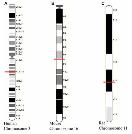 Fig. 1 Location of growth-associated protein (GAP)-43 gene on chromosomes of different species.1
Key structural properties of GAP43:
- Highly flexible irregular coiled conformation
- The N-terminal membrane anchoring region contains palmitoylation modification sites
- The central region contains a PKC-specific phosphorylation site (Ser41)
- Lacks a typical hydrophobic core and is stabilized by membrane binding
- Rich in alanine and proline residues
- Phosphorylation status regulates the ability to bind to actin
Functions of GAP43
The main function of GAP43 is to regulate the movement of nerve growth cones and synaptic plasticity. In addition, it also plays a role in axon guidance, nerve regeneration, and synaptic vesicle cycling.
| Function | Description |
| Axonal guidance | GAP43 is enriched locally in the growth cone membrane, mediating the coupling of extracellular guidance signals with the rearrangement of the actin cytoskeleton. |
| Synaptic plasticity | Involved in the induction of long-term potentiation, regulating the efficiency of neurotransmitter release at the presynaptic terminal. |
| Neural regeneration | Expressed significantly upregulated after peripheral nerve injury, promoting axonal sprouting and targeted reconnection. |
| Cytoskeleton regulation | The phosphorylation state determines its binding ability to the submembrane actin, affecting the turning of the growth cone. |
| Learning and memory related | Highly expressed in hippocampus and cortical brain regions, closely related to the formation of spatial memory. |
The expression of GAP43 is regulated by developmental stages. It is highly expressed during the embryonic period and is downregulated in most brain regions in adulthood. Only in plastic regions such as the olfactory bulb and hippocampus does it maintain a relatively high level.
Applications of GAP43 and GAP43 Antibody in Literature
1. Holahan, Matthew R. "A shift from a pivotal to supporting role for the growth-associated protein (GAP-43) in the coordination of axonal structural and functional plasticity." Frontiers in Cellular Neuroscience 11 (2017): 266. https://doi.org/10.3389/fncel.2017.00266
The article indicates that GAP-43, as a key regulatory protein for axon growth and plasticity, participates in synaptic function regulation through its post-transcriptional modifications and protein interactions. Although it is not a sufficient condition for neural regeneration, the related research has deepened our understanding of the biochemical mechanism of axon plasticity.
2. Maroto, Irene B., et al. "GAP43 located on corticostriatal terminals restrains novelty-induced hyperactivity in mice." Journal of Neuroscience 44.39 (2024). https://doi.org/10.1523/JNEUROSCI.0701-24.2024
The research found that in mice with specific deletion of the GAP-43 protein in glutamatergic neurons of the telencephalon, the excitatory synaptic transmission in the cortical striatal circuit would be enhanced, leading to excessive active behaviors in new environments. This suggests that GAP-43 plays a crucial regulatory role in this behavior circuit.
3. Yang, Mingming, et al. "Cryptic Splicing of GAP43 mRNA is a Novel Hallmark of TDP‐43‐Associated ALS and AD." Advanced Science (2025): e12054. https://doi.org/10.1002/advs.202412054
The research has found that the TDP-43 protein regulates the expression of GAP43 precursor RNA by binding to it. Abnormal function of TDP-43 can induce covert splicing of GAP43, resulting in a decrease in its protein level, thereby impairing axonal regeneration. This discovery reveals a new mechanism of neuronal degeneration in ALS and AD.
4. Zhang, Fanrong, et al. "GAP43, a novel metastasis promoter in non-small cell lung cancer." Journal of translational medicine 16.1 (2018): 310. https://doi.org/10.1186/s12967-018-1682-5
The study found that GAP43 is highly expressed in non-small cell lung cancer and can promote tumor cell migration and brain metastasis by activating the Rac1/F-actin pathway. It can be used as an independent biomarker for predicting the risk of brain metastasis in patients.
5. Li, Pingjiang, et al. "lncRNA LOC100911717-targeting GAP43-mediated sympathetic remodeling after myocardial infarction in rats." Frontiers in cardiovascular medicine 9 (2023): 1019435. https://doi.org/10.3389/fcvm.2022.1019435
The research has found that the elevated lncRNA LOC100911717 after myocardial infarction can promote sympathetic nerve remodeling and ventricular arrhythmias by up-regulating the expression of GAP43. Its silencing can alleviate these pathological changes, providing a new target for the treatment of arrhythmia.
Creative Biolabs: GAP43 Antibodies for Research
Creative Biolabs specializes in the production of high-quality GAP43 antibodies for research and industrial applications. Our portfolio includes monoclonal and polyclonal antibodies tailored for ELISA, Flow Cytometry, Western blot, immunohistochemistry, and other diagnostic methodologies.
- Custom GAP43 Antibody Development: Tailor-made solutions to meet specific research requirements.
- Bulk Production: Large-scale antibody manufacturing for industry partners.
- Technical Support: Expert consultation for protocol optimization and troubleshooting.
- Aliquoting Services: Conveniently sized aliquots for long-term storage and consistent experimental outcomes.
For more details on our GAP43 antibodies, custom preparations, or technical support, contact us at info@creative-biolabs.com.
Reference
- Holahan, Matthew R. "A shift from a pivotal to supporting role for the growth-associated protein (GAP-43) in the coordination of axonal structural and functional plasticity." Frontiers in Cellular Neuroscience 11 (2017): 266. Distributed under Open Access license CC BY 4.0, without modification. https://doi.org/10.3389/fncel.2017.00266
Anti-GAP43 antibodies
 Loading...


Hot products 
-
Mouse Anti-FYN Recombinant Antibody (10) (CBMAB-S6332-CQ)

-
Rabbit Anti-CAMK2A Recombinant Antibody (BA0032) (CBMAB-0137CQ)

-
Mouse Anti-GLP1R Recombinant Antibody (4F3) (CBMAB-G0521-LY)

-
Mouse Anti-CD46 Recombinant Antibody (CBFYC-0076) (CBMAB-C0085-FY)

-
Mouse Anti-ADV Recombinant Antibody (V2-503423) (CBMAB-V208-1364-FY)

-
Mouse Anti-CSPG4 Recombinant Antibody (CBFYM-1050) (CBMAB-M1203-FY)

-
Mouse Anti-C5b-9 Recombinant Antibody (aE11) (CBMAB-AO138LY)

-
Mouse Anti-GFAP Recombinant Antibody (5) (CBMAB-G0346-LY)

-
Mouse Anti-ACTB Recombinant Antibody (V2-179553) (CBMAB-A0870-YC)

-
Mouse Anti-DLL4 Recombinant Antibody (D1090) (CBMAB-D1090-YC)

-
Mouse Anti-DISP2 Monoclonal Antibody (F66A4B1) (CBMAB-1112CQ)

-
Mouse Anti-CFL1 Recombinant Antibody (CBFYC-1771) (CBMAB-C1833-FY)

-
Mouse Anti-CD33 Recombinant Antibody (6C5/2) (CBMAB-C8126-LY)

-
Rabbit Anti-ADRA1A Recombinant Antibody (V2-12532) (CBMAB-1022-CN)

-
Mouse Anti-ADAM29 Recombinant Antibody (V2-179787) (CBMAB-A1149-YC)

-
Rabbit Anti-B2M Recombinant Antibody (CBYY-0059) (CBMAB-0059-YY)

-
Mouse Anti-BAX Recombinant Antibody (CBYY-0216) (CBMAB-0217-YY)

-
Mouse Anti-AMOT Recombinant Antibody (CBYC-A564) (CBMAB-A2552-YC)

-
Mouse Anti-CTNND1 Recombinant Antibody (CBFYC-2414) (CBMAB-C2487-FY)

-
Mouse Anti-AOC3 Recombinant Antibody (CBYY-0014) (CBMAB-0014-YY)

- AActivation
- AGAgonist
- APApoptosis
- BBlocking
- BABioassay
- BIBioimaging
- CImmunohistochemistry-Frozen Sections
- CIChromatin Immunoprecipitation
- CTCytotoxicity
- CSCostimulation
- DDepletion
- DBDot Blot
- EELISA
- ECELISA(Cap)
- EDELISA(Det)
- ESELISpot
- EMElectron Microscopy
- FFlow Cytometry
- FNFunction Assay
- GSGel Supershift
- IInhibition
- IAEnzyme Immunoassay
- ICImmunocytochemistry
- IDImmunodiffusion
- IEImmunoelectrophoresis
- IFImmunofluorescence
- IGImmunochromatography
- IHImmunohistochemistry
- IMImmunomicroscopy
- IOImmunoassay
- IPImmunoprecipitation
- ISIntracellular Staining for Flow Cytometry
- LALuminex Assay
- LFLateral Flow Immunoassay
- MMicroarray
- MCMass Cytometry/CyTOF
- MDMeDIP
- MSElectrophoretic Mobility Shift Assay
- NNeutralization
- PImmunohistologyp-Paraffin Sections
- PAPeptide Array
- PEPeptide ELISA
- PLProximity Ligation Assay
- RRadioimmunoassay
- SStimulation
- SESandwich ELISA
- SHIn situ hybridization
- TCTissue Culture
- WBWestern Blot
