GCGR Antibodies
Background
GCGR (glucagon receptor) is a G protein-coupled receptor mainly distributed on the cell membranes of tissues such as the liver and kidneys. This receptor can specifically bind to glucagon, thereby activating intracellular signaling pathways, promoting glycogenolysis and glucose production, and thus playing a core regulatory role in maintaining blood glucose homeostasis. The functional abnormalities of GCGR are closely related to metabolic diseases such as type 2 diabetes and obesity. Since its coding gene was cloned and identified in the 1990s, this receptor has become a key target in the development of diabetes drugs. Its complex transmembrane structure and dynamic signal transduction mechanism have been continuously and deeply studied, greatly promoting the understanding of the molecular mechanisms of G protein-coupled receptor biology and metabolic regulation.
Structure of GCGR
The glucagon receptor encoded by the GCGR gene is a G protein-coupled receptor with a molecular weight of approximately 55 kDa. This molecular weight may slightly fluctuate in different cell expression systems due to the degree of glycosylation modification.
| Species | Human | Mouse | Rat |
| Molecular Weight (kDa) | ~55 | ~54 | ~55 |
| Primary Structural Differences | Classical seven-fold transmembrane structure with specific glycosylation sites at the N-terminus | Across the membrane area highly conservative, subtle differences cell inner sequence | High homology with human receptors and similar signal transduction function |
This receptor is composed of approximately 477 amino acids, and its primary structure presents a typical GPCR sequence pattern, including an extracellular N-terminal domain, seven hydrophobic transmembrane α -helical regions (TM1-TM7), and an intracellular C-terminal tail. The core of its protein structure lies in the ligand-binding pocket formed by the transmembrane helix, as well as the crucial intracellular loop (especially the third intracellular loop) for coupling G proteins. The secondary structure is dominated by transmembrane α -helices, which are connected by annular regions. Its functional activity depends on a characteristic structural motif: amino acid residues located near TM5 and TM6 together form the binding site of glucagon, while the intracellular region contains the conserved motif necessary for interaction with Gs proteins. The activation of receptors triggers conformational changes, which in turn activate downstream blood glucose regulatory signaling pathways.
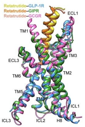 Fig. 1 Structural comparison of retatrutide–GLP-1R, retatrutide–GIPR and retatrutide–GCGR.1
Key structural properties of GCGR:
- Classic seven-transmembrane α helix structure
- Hydrophobic ligand-binding pockets formed by transmembrane helices
- The intracellular loop and the C-terminal region contain multiple key motifs that couple with G proteins (mainly Gs)
- Conserved polar amino acid networks and disulfide bonds
Functions of GCGR
The main function of the GCGR (glucagon receptor) gene is to mediate the signaling of glucagon, thereby regulating blood glucose homeostasis. However, this receptor is also involved in a variety of other physiological and pathological processes, including energy metabolism, insulin secretion regulation, and liver lipid metabolism, etc.
| Function | Description |
| Elevated blood sugar | After the receptor is activated, it activates adenylate cyclase through Gs protein, increasing the intracellular cAMP level, promoting liver glycogenolysis and gluconeogenesis, and thereby raising blood sugar. |
| Metabolic regulation | In addition to sugar metabolism, it also participates in regulating lipid breakdown and ketogenesis, influencing the balance of energy metabolism throughout the body. |
| Regulation of insulin secretion | In pancreatic β cells, the activation of GCGR can regulate insulin secretion stimulated by glucose at specific stages. |
| Liver protection and regeneration | Studies have shown that the glucagon-GCGR signaling pathway is involved in regulating hepatocyte proliferation and liver regeneration processes. |
| Disease association | Its hyperfunction is associated with type 2 diabetes and hyperglycemia, while functional inhibition or antagonism is regarded as a potential therapeutic strategy. |
The signal transduction dynamics of GCGR exhibit a rapid and tunable saturation response mode, which contrasts sharply with the continuous oxygen-binding characteristics of myoglobin as a reserve protein, demonstrating its immediate sensing and regulatory role in maintaining dynamic blood glucose balance.
Applications of GCGR and GCGR Antibody in Literature
1. Zimmermann, Tina, et al. "BI 456906: Discovery and preclinical pharmacology of a novel GCGR/GLP-1R dual agonist with robust anti-obesity efficacy." Molecular metabolism 66 (2022): 101633.https://doi.org/10.1016/j.molmet.2022.101633
The article indicates that BI 456906 is a novel dual agonist of GCGR/GLP-1R, which achieves potent weight loss by increasing energy expenditure and reducing food intake. Its efficacy is superior to that of a single GLP-1R agonist and supports administration once a week.
2. Li, Wenzhuo, et al. "Structural insights into the triple agonism at GLP-1R, GIPR and GCGR manifested by retatrutide." Cell Discovery 10.1 (2024): 77.https://doi.org/10.1038/s41421-024-00700-0
The article indicates that the novel triple agonist retatrutide can simultaneously act on GLP-1R, GIPR and GCGR. By activating GCGR, it increases energy expenditure and has shown superior weight loss and blood sugar control effects compared to existing drugs in clinical trials for obesity and type 2 diabetes.
3. Zheng, Yi, et al. "CD9 counteracts liver steatosis and mediates GCGR agonist hepatic effects." Advanced Science 11.29 (2024): 2400819. https://doi.org/10.1002/advs.202400819
This article reveals that protein CD9 mediates the mechanism by which GCGR agonists improve liver steatosis. CD9 in the liver regulates the expression of CFD by ubiquitinating and degrading FLI1, thereby influencing fatty acid metabolism. Targeting CD9 may become a new strategy for treating fatty liver.
4. Cui, Xiaona, et al. "Pancreatic alpha cell glucagon–liver FGF21 axis regulates beta cell regeneration in a mouse model of type 2 diabetes." Diabetologia 66.3 (2023): 535-550. https://doi.org/10.1007/s00125-022-05822-2
Studies have shown that in a mouse model of type 2 diabetes, antagonizing GCGR can improve hyperglycemia and promote the regeneration of pancreatic β cells. This effect is mediated by liver-derived FGF21, enhancing the expression of genes related to β -cell proliferation and function.
5. Puszkarska, Anna M., et al. "Machine learning designs new GCGR/GLP-1R dual agonists with enhanced biological potency." Nature Chemistry 16.9 (2024): 1436-1444. https://doi.org/10.1038/s41557-024-01532-x
In this study, a deep learning model was utilized to successfully design multiple novel dual-target peptide variants based on the GCGR/GLP-1R dual activation activity data of known peptide sequences. Among them, the agonistic activities of some variants on both receptors were significantly enhanced, with the highest reaching seven times that of the best sequence in the training set.
Creative Biolabs: GCGR Antibodies for Research
Creative Biolabs specializes in the production of high-quality GCGR antibodies for research and industrial applications. Our portfolio includes monoclonal antibodies tailored for ELISA, Flow Cytometry, Western blot, immunohistochemistry, and other diagnostic methodologies.
- Custom GCGR Antibody Development: Tailor-made solutions to meet specific research requirements.
- Bulk Production: Large-scale antibody manufacturing for industry partners.
- Technical Support: Expert consultation for protocol optimization and troubleshooting.
- Aliquoting Services: Conveniently sized aliquots for long-term storage and consistent experimental outcomes.
For more details on our GCGR antibodies, custom preparations, or technical support, contact us at email.
Reference
- Li, Wenzhuo, et al. "Structural insights into the triple agonism at GLP-1R, GIPR and GCGR manifested by retatrutide." Cell Discovery 10.1 (2024): 77. https://doi.org/10.1038/s41421-024-00700-0
Anti-GCGR antibodies
 Loading...


Hot products 
-
Mouse Anti-BIRC7 Recombinant Antibody (88C570) (CBMAB-L0261-YJ)

-
Mouse Anti-BCL6 Recombinant Antibody (CBYY-0435) (CBMAB-0437-YY)

-
Mouse Anti-AZGP1 Recombinant Antibody (CBWJZ-007) (CBMAB-Z0012-WJ)

-
Mouse Anti-GDF5 Recombinant Antibody (1F4) (CBMAB-G2740-LY)

-
Mouse Anti-CD8 Recombinant Antibody (C1083) (CBMAB-C1083-LY)

-
Mouse Anti-FOXA3 Recombinant Antibody (2A9) (CBMAB-0377-YC)

-
Mouse Anti-ADGRE5 Recombinant Antibody (V2-360335) (CBMAB-C2088-CQ)

-
Rabbit Anti-ENO2 Recombinant Antibody (BA0013) (CBMAB-0272CQ)

-
Mouse Anti-G6PD Recombinant Antibody (13B331) (CBMAB-G1553-LY)

-
Mouse Anti-ARSA Recombinant Antibody (CBYC-A799) (CBMAB-A3679-YC)

-
Mouse Anti-CD24 Recombinant Antibody (ALB9) (CBMAB-0176CQ)

-
Mouse Anti-AQP2 Recombinant Antibody (E-2) (CBMAB-A3358-YC)

-
Mouse Anti-ADAM12 Recombinant Antibody (V2-179752) (CBMAB-A1114-YC)

-
Mouse Anti-CARTPT Recombinant Antibody (113612) (CBMAB-C2450-LY)

-
Rat Anti-CCR2 Recombinant Antibody (475301) (CBMAB-C1338-LY)

-
Mouse Anti-CEMIP Recombinant Antibody (3C12) (CBMAB-K0296-LY)

-
Mouse Anti-FN1 Monoclonal Antibody (71) (CBMAB-1241CQ)

-
Mouse Anti-CCN2 Recombinant Antibody (CBFYC-2383) (CBMAB-C2456-FY)

-
Mouse Anti-EMP3 Recombinant Antibody (CBFYE-0100) (CBMAB-E0207-FY)

-
Mouse Anti-ASB9 Recombinant Antibody (1D8) (CBMAB-A0529-LY)

- AActivation
- AGAgonist
- APApoptosis
- BBlocking
- BABioassay
- BIBioimaging
- CImmunohistochemistry-Frozen Sections
- CIChromatin Immunoprecipitation
- CTCytotoxicity
- CSCostimulation
- DDepletion
- DBDot Blot
- EELISA
- ECELISA(Cap)
- EDELISA(Det)
- ESELISpot
- EMElectron Microscopy
- FFlow Cytometry
- FNFunction Assay
- GSGel Supershift
- IInhibition
- IAEnzyme Immunoassay
- ICImmunocytochemistry
- IDImmunodiffusion
- IEImmunoelectrophoresis
- IFImmunofluorescence
- IGImmunochromatography
- IHImmunohistochemistry
- IMImmunomicroscopy
- IOImmunoassay
- IPImmunoprecipitation
- ISIntracellular Staining for Flow Cytometry
- LALuminex Assay
- LFLateral Flow Immunoassay
- MMicroarray
- MCMass Cytometry/CyTOF
- MDMeDIP
- MSElectrophoretic Mobility Shift Assay
- NNeutralization
- PImmunohistologyp-Paraffin Sections
- PAPeptide Array
- PEPeptide ELISA
- PLProximity Ligation Assay
- RRadioimmunoassay
- SStimulation
- SESandwich ELISA
- SHIn situ hybridization
- TCTissue Culture
- WBWestern Blot
