MKI67 Antibodies
Background
The MKI67 gene encodes a proliferation marker protein present in the cell nucleus and is widely distributed in actively dividing cells. This protein participates in cell cycle regulation, ribosomal RNA synthesis and mitosis process through interaction with chromosomes, and its expression level directly reflects the proliferation status of cells. Due to its significantly high expression in tumor tissues, MKI67 has become a key indicator for evaluating the malignancy of tumors in clinical pathological diagnosis. This gene was first discovered by scholars such as Schluter in 1983 during their research on Hodgkin's lymphoma. Its name is derived from the University of Kiel in Germany and the discovery number. As a classic molecule in cell dynamics research, MKI67 not only provides an important basis for tumor prognosis judgment, but also deepens people's understanding of the cell cycle regulation mechanism, becoming a key bridge connecting basic research and clinical practice.
Structure of MKI67
MKI67 is a large nucleoprotein with a molecular weight of approximately 359 kDa. This protein is encoded by a gene of up to 11,423 bases, and its molecular weight can vary significantly due to the differences in the splicing methods of the transcript among different species.
| Species | Human | Mouse | Rat | Bovine |
| Molecular Weight (kb) | About 40 | About 38 | About 39 | About 41 |
| Primary Structural Differences | Contains multiple conserved domains | Structure is similar to human height | There are short fragment differences | Relatively high conservatism |
| Protein isomers | Two main types | Two main types | Two main types | Two main types |
This protein contains multiple characteristic domains, among which the most important one is the "Ki67 domain" encoded by exon 13, which forms a typical coil-coil conformation. The N-terminal and C-terminal regions respectively contain highly conserved PP1 phosphorylation sites and tandem repeat sequences. These repeat units mediate the interaction between the protein and chromatin by forming specific spatial conformations. Its secondary structure is mainly composed of α -helices, which are assembled into a typical bull-shaped form through flexible junction regions, enabling it to dynamically bind to the chromosome surface during cell division.
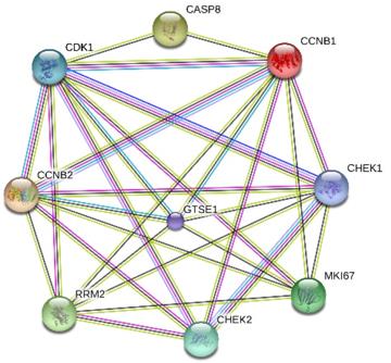 Fig. 1 Interactions between MKI67 and eight genes related to P53 signaling pathway.1
Key structural properties of MKI67:
- Contains large coiled helical domains
- Modules with tandem repeats are used for chromatin binding
- Contains multiple PP1 phosphorylation sites to regulate cell cycle
- Form a bull-shaped dimer structure to scan the surface of chromosomes
Functions of MKI67
The core function of the MKI67 gene is to serve as a marker for cell proliferation. However, it is also directly involved in a variety of key cell cycle processes, including the maintenance of chromosomal organization and the regulation of mitosis.
| Function | Description |
| Proliferation marker | In active dividing cells throughout the cell cycle phase (G1, S, G2, M) in the continued to express, to provide clinical key proliferation activity index.. |
| Chromosomal organization | By interacting with heterochromatin and nucleolar tissue regions, it helps maintain the structure and spatial configuration of nucleoli during the intermitotic phase. |
| Mitotic regulation | During the cell division phase, its protein products form the "Ki67 coat" surrounding the chromosome, and the physical barrier nuclear membrane reforms at the wrong position. |
| Ribosome biosynthesis | Involved in regulation of ribosomal RNA synthesis process, indirect influence the growth and proliferation of cells. |
| Prognosis assessment | In tumor tissues, its expression level is closely related to the degree of malignancy and the risk of metastasis, and it is a core assessment parameter in pathological diagnosis. |
Unlike cyclins that only function at specific cycle stages, the expression of MKI67 is continuous throughout the entire cycle. This unique dynamic pattern makes it a "molecular ruler" for precisely quantifying the proportion of proliferating cells in complex tissues.
Applications of MKI67 and MKI67 Antibody in Literature
1. Saeed, Muhammad Muddasar, et al. "RACGAP1 and MKI67 are potential prognostic biomarker in hepatocellular carcinoma caused by HBV/HCV via lactylation." Frontiers in Oncology 15 (2025): 1537084. https://doi.org/10.3389/fonc.2025.1537084
This study found through sequencing that MKI67 is a key gene for pulmonary arterial hypertension. Experiments have confirmed that inhibiting MKI67 can significantly reduce the proliferation and migration of pulmonary artery smooth muscle cells, indicating its potential as a new target for the diagnosis and treatment of pulmonary arterial hypertension.
2. Wu, Shi-yi, et al. "Correlation of MKI67 with prognosis, immune infiltration, and T cell exhaustion in hepatocellular carcinoma." BMC gastroenterology 21.1 (2021): 416. https://doi.org/10.1186/s12876-021-01984-2
Research has found that MKI67 is a key gene regulating the proliferation and migration of pulmonary artery smooth muscle cells. Inhibiting its expression can slow down the lesion process, indicating that MKI67 is expected to become a new diagnostic and therapeutic target for pulmonary arterial hypertension.
3. Xiong, Dan-dan, et al. "Ki-67/MKI67 as a predictive biomarker for clinical outcome in gastric cancer patients: an updated meta-analysis and systematic review involving 53 studies and 7078 patients." Journal of Cancer 10.22 (2019): 5339. https://doi.org/10.7150/jca.30074
Studies have found that gastric cancer patients with high expression of Ki-67/MKI67 have a poorer overall survival period, and it is significantly associated with adverse features such as advanced tumor and lymph node metastasis. It can serve as a potential indicator for prognosis prediction and identification of high-risk cases.
4. Pan, Xiaolan, et al. "A comprehensive analysis of the prognostic value, expression characteristics and immune correlation of MKI67 in cancers." Frontiers in Immunology 16 (2025): 1531708. https://doi.org/10.3389/fimmu.2025.1531708
Pan-cancer analysis shows that MKI67 is highly expressed in various cancers and is significantly associated with poor prognosis in patients with lung adenocarcinoma and liver cancer. It also involves pathways such as the cell cycle and p53, and has important clinical prognostic guidance value.
5. Zhou, Huiling, et al. "MKI67 as a potential diagnostic biomarker in pulmonary hypertension." Frontiers in Pediatrics 10 (2022): 1016889. https://doi.org/10.3389/fped.2022.1016889
Research has found that MKI67 is a key gene regulating the proliferation and migration of pulmonary artery smooth muscle cells. Inhibiting its expression can slow down the progression of the disease and is expected to become a new target for the diagnosis and treatment of pulmonary arterial hypertension.
Creative Biolabs: MKI67 Antibodies for Research
Creative Biolabs specializes in the production of high-quality MKI67 antibodies for research and industrial applications. Our portfolio includes monoclonal antibodies tailored for ELISA, Flow Cytometry, Western blot, immunohistochemistry, and other diagnostic methodologies.
- Custom MKI67 Antibody Development: Tailor-made solutions to meet specific research requirements.
- Bulk Production: Large-scale antibody manufacturing for industry partners.
- Technical Support: Expert consultation for protocol optimization and troubleshooting.
- Aliquoting Services: Conveniently sized aliquots for long-term storage and consistent experimental outcomes.
For more details on our MKI67 antibodies, custom preparations, or technical support, contact us at email.
Reference
- Xiong, Dan-dan, et al. "Ki-67/MKI67 as a predictive biomarker for clinical outcome in gastric cancer patients: an updated meta-analysis and systematic review involving 53 studies and 7078 patients." Journal of Cancer 10.22 (2019): 5339. https://doi.org/10.7150/jca.30074
Anti-MKI67 antibodies
 Loading...


Hot products 
-
Mouse Anti-F11R Recombinant Antibody (402) (CBMAB-0026-WJ)

-
Mouse Anti-ADGRL2 Recombinant Antibody (V2-58519) (CBMAB-L0166-YJ)

-
Mouse Anti-CSPG4 Recombinant Antibody (CBFYM-1050) (CBMAB-M1203-FY)

-
Mouse Anti-CD2AP Recombinant Antibody (BR083) (CBMAB-BR083LY)

-
Mouse Anti-BCL6 Recombinant Antibody (CBYY-0442) (CBMAB-0445-YY)

-
Mouse Anti-BPGM Recombinant Antibody (CBYY-1806) (CBMAB-2155-YY)

-
Mouse Anti-CARTPT Recombinant Antibody (113612) (CBMAB-C2450-LY)

-
Mouse Anti-GGT1 Recombinant Antibody (1F9) (CBMAB-G3273-LY)

-
Mouse Anti-ELAVL4 Recombinant Antibody (6B9) (CBMAB-1132-YC)

-
Mouse Anti-C5b-9 Recombinant Antibody (aE11) (CBMAB-AO138LY)

-
Mouse Anti-C5AR1 Recombinant Antibody (R63) (CBMAB-C9553-LY)

-
Mouse Anti-AKT1 (Phosphorylated S473) Recombinant Antibody (V2-505430) (PTM-CBMAB-0067LY)

-
Mouse Anti-AAV9 Recombinant Antibody (V2-634029) (CBMAB-AP023LY)

-
Mouse Anti-4-Hydroxynonenal Recombinant Antibody (V2-502280) (CBMAB-C1055-CN)

-
Mouse Anti-CD83 Recombinant Antibody (HB15) (CBMAB-C1765-CQ)

-
Mouse Anti-ATP1B1 Recombinant Antibody (E4) (CBMAB-0463-LY)

-
Mouse Anti-DLL4 Recombinant Antibody (D1090) (CBMAB-D1090-YC)

-
Mouse Anti-ASH1L Monoclonal Antibody (ASH5H03) (CBMAB-1372-YC)

-
Mouse Anti-DLC1 Recombinant Antibody (D1009) (CBMAB-D1009-YC)

-
Mouse Anti-ANXA7 Recombinant Antibody (A-1) (CBMAB-A2941-YC)

- AActivation
- AGAgonist
- APApoptosis
- BBlocking
- BABioassay
- BIBioimaging
- CImmunohistochemistry-Frozen Sections
- CIChromatin Immunoprecipitation
- CTCytotoxicity
- CSCostimulation
- DDepletion
- DBDot Blot
- EELISA
- ECELISA(Cap)
- EDELISA(Det)
- ESELISpot
- EMElectron Microscopy
- FFlow Cytometry
- FNFunction Assay
- GSGel Supershift
- IInhibition
- IAEnzyme Immunoassay
- ICImmunocytochemistry
- IDImmunodiffusion
- IEImmunoelectrophoresis
- IFImmunofluorescence
- IGImmunochromatography
- IHImmunohistochemistry
- IMImmunomicroscopy
- IOImmunoassay
- IPImmunoprecipitation
- ISIntracellular Staining for Flow Cytometry
- LALuminex Assay
- LFLateral Flow Immunoassay
- MMicroarray
- MCMass Cytometry/CyTOF
- MDMeDIP
- MSElectrophoretic Mobility Shift Assay
- NNeutralization
- PImmunohistologyp-Paraffin Sections
- PAPeptide Array
- PEPeptide ELISA
- PLProximity Ligation Assay
- RRadioimmunoassay
- SStimulation
- SESandwich ELISA
- SHIn situ hybridization
- TCTissue Culture
- WBWestern Blot
