NFIC Antibodies
Background
The NFIC gene encodes Nuclear Factor I/C, which is an important transcriptional regulatory factor widely expressed in vertebrates. This protein regulates the transcription process of downstream target genes by recognizing specific DNA sequences, and plays a particularly crucial role in organ development, cell differentiation, and maintaining metabolic balance. In the formation of teeth and bones, NFIC has been proven to regulate root development by influencing the differentiation of odontoblasts, and its mutations can lead to abnormal development of related tissues. Since its discovery in the 1980s, this gene has become an important model in developmental biology and disease mechanism research due to its core position in the tissue-specific transcriptional regulatory network, providing a key theoretical basis for understanding the role of transcription factors in cell fate determination and morphogenesis.
Structure of NFIC
The nuclear factor I/C (NFIC) encoded by the NFIC gene is a transcriptional regulatory protein with a molecular weight of approximately 55-65 kDa. Its precise molecular weight may vary due to different splicing isomers and post-translational modifications such as phosphorylation. This protein contains a highly conserved DNA-binding domain that can specifically recognize and bind to the TTGGCNNNNNGCCAA sequence in the promoter region of the target gene. The basic amino acid region in its primary structure is responsible for nuclear localization, while the proline-rich region mediates transcriptional activation function. NFIC proteins function by forming homologous or heterodimers. The dimerization domain ensures their high affinity for DNA binding, thereby precisely regulating the gene expression network related to cell differentiation and organ development (such as teeth and the brain).
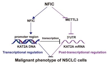 Fig. 1 NFIC suppresses NSCLC progression via the METTL3/KAT2A axis.1
Key structural properties of NFIC:
- Conserved DNA-binding domains (including helical - angular - helical mods)
- Dimerization domains mediate protein oligomerization
- Nuclear localization signals guide proteins to enter the nucleus
Functions of NFIC
The core function of the NFIC gene-encoded protein is to regulate gene transcription to maintain the balance of cell differentiation. It participates in multiple biological processes simultaneously, including organ development and metabolic regulation.
| Function | Description |
| Transcriptional regulation | By identifying specific DNA sequences to initiate the transcription of target genes, it guides the directional differentiation of cells. |
| Organ development | Regulate the morphogenesis and tissue-specific formation of organs such as teeth and bones. |
| Metabolic homeostasis | Affect the glucolipid metabolism related gene expression, maintain energy balance. |
| Cell cycle regulation | Inhibit abnormal proliferation and promote the normal differentiation process of cells. |
| Damage repair | Activate the expression network of genes related to repair after tissue damage. |
This transcription factor achieves precise regulation of downstream genes through a synergistic mechanism: its DNA-binding domain recognizes target sequences with high affinity, while the dimerization domain regulates transcriptional activity by forming specific spatial conformations. This dual mechanism ensures its core regulatory position in development and metabolic processes.
Applications of NFIC and NFIC Antibody in Literature
1. Cobo, Isidoro, et al. "NFIC regulates ribosomal biology and ER stress in pancreatic acinar cells and restrains PDAC initiation." Nature Communications 14.1 (2023): 3761.https://doi.org/10.1038/s41467-023-39291-x
Research has found that the transcription factor NFIC interacts with NR5A2 to regulate the differentiation and function of pancreatic acinar cells and alleviate endoplasmic reticulum stress. Deletion of NFIC can impede the repair of pancreatic injury and promote the development of precancerous lesions driven by Kras mutations.
2. Huang, Huimin, et al. "Adipose-derived stem cell exosome NFIC improves diabetic foot ulcers by regulating miR-204-3p/HIPK2." Journal of orthopaedic surgery and research 18.1 (2023): 687. https://doi.org/10.1186/s13018-023-04165-x
Research has found that adipocytes can deliver the transcription factor NFIC through exosomes, thereby activating miR-204-3p and inhibiting its target gene HIPK2, which promotes the repair of vascular endothelial cells in a high-glucose environment and provides a new approach for the treatment of diabetic foot ulcers.
3. Shi, Kesong, et al. "NFIC mediates m6A mRNA methylation to orchestrate transcriptional and post-transcriptional regulation to represses malignant phenotype of non-small cell lung cancer cells." Cancer Cell International 24.1 (2024): 223. https://doi.org/10.1186/s12935-024-03414-1
Research has found that the transcription factor NFIC inhibits the malignant progression of non-small cell lung cancer by directly suppressing the METTL3 and KAT2A genes and reducing the m6A modification level. NFIC is lowly expressed in cancer tissues and exerts a tumor suppressor effect.
4. Polcaro, Giovanna, et al. "rs822336 binding to C/EBPβ and NFIC modulates induction of PD-L1 expression and predicts anti-PD-1/PD-L1 therapy in advanced NSCLC." Molecular Cancer 23.1 (2024): 63.https://doi.org/10.1186/s12943-024-01976-2
Research has found that the PD-L1 gene locus rs822336 can be used as a biomarker for predicting the efficacy of immunotherapy in non-small cell lung cancer. This site affects the expression level of PD-L1 by regulating the competitive binding of the transcription factor C/EBPβ to NFIC, thereby determining the response to immunotherapy.
5. Jiang, Shuang, et al. "USP34 regulates tooth root morphogenesis by stabilizing NFIC." International Journal of Oral Science 13.1 (2021): 7. https://doi.org/10.1038/s41368-021-00114-8
Research has found that the deubiquitinating enzyme USP34 regulates root development by stabilizing the transcription factor NFIC. The absence of USP34 can lead to the obstruction of odontogenic differentiation of dental mesenchymal cells, resulting in developmental defects such as shortened tooth roots and thinner dentin.
Creative Biolabs: NFIC Antibodies for Research
Creative Biolabs specializes in the production of high-quality NFIC antibodies for research and industrial applications. Our portfolio includes monoclonal antibodies tailored for ELISA, Flow Cytometry, Western blot, immunohistochemistry, and other diagnostic methodologies.
- Custom NFIC Antibody Development: Tailor-made solutions to meet specific research requirements.
- Bulk Production: Large-scale antibody manufacturing for industry partners.
- Technical Support: Expert consultation for protocol optimization and troubleshooting.
- Aliquoting Services: Conveniently sized aliquots for long-term storage and consistent experimental outcomes.
For more details on our NFIC antibodies, custom preparations, or technical support, contact us at email.
Reference
- Shi, Kesong, et al. "NFIC mediates m6A mRNA methylation to orchestrate transcriptional and post-transcriptional regulation to represses malignant phenotype of non-small cell lung cancer cells." Cancer Cell International 24.1 (2024): 223. https://doi.org/10.1186/s12935-024-03414-1
Anti-NFIC antibodies
 Loading...


Hot products 
-
Rat Anti-CD300A Recombinant Antibody (172224) (CBMAB-C0423-LY)

-
Mouse Anti-DDC Recombinant Antibody (8E8) (CBMAB-0992-YC)

-
Mouse Anti-DHFR Recombinant Antibody (D0821) (CBMAB-D0821-YC)

-
Mouse Anti-ESR1 Recombinant Antibody (Y31) (CBMAB-1208-YC)

-
Human Anti-SARS-CoV-2 Spike Recombinant Antibody (CR3022) (CBMAB-CR014LY)

-
Mouse Anti-CD33 Recombinant Antibody (6C5/2) (CBMAB-C8126-LY)

-
Rat Anti-4-1BB Recombinant Antibody (V2-1558) (CBMAB-0953-LY)

-
Mouse Anti-AHCYL1 Recombinant Antibody (V2-180270) (CBMAB-A1703-YC)

-
Mouse Anti-ADAM29 Recombinant Antibody (V2-179787) (CBMAB-A1149-YC)

-
Mouse Anti-BHMT Recombinant Antibody (CBYY-0547) (CBMAB-0550-YY)

-
Mouse Anti-APOH Recombinant Antibody (4D9A4) (CBMAB-A3249-YC)

-
Mouse Anti-CCT6A/B Recombinant Antibody (CBXC-0168) (CBMAB-C5570-CQ)

-
Mouse Anti-DMD Recombinant Antibody (D1190) (CBMAB-D1190-YC)

-
Mouse Anti-ADV Recombinant Antibody (V2-503423) (CBMAB-V208-1364-FY)

-
Rabbit Anti-ENO2 Recombinant Antibody (BA0013) (CBMAB-0272CQ)

-
Mouse Anti-ASB9 Recombinant Antibody (1D8) (CBMAB-A0529-LY)

-
Mouse Anti-CD8 Recombinant Antibody (C1083) (CBMAB-C1083-LY)

-
Rat Anti-CD34 Recombinant Antibody (MEC 14.7) (CBMAB-C10196-LY)

-
Mouse Anti-ALDOA Recombinant Antibody (A2) (CBMAB-A2316-YC)

-
Mouse Anti-AKT1/AKT2/AKT3 (Phosphorylated T308, T309, T305) Recombinant Antibody (V2-443454) (PTM-CBMAB-0030YC)

- AActivation
- AGAgonist
- APApoptosis
- BBlocking
- BABioassay
- BIBioimaging
- CImmunohistochemistry-Frozen Sections
- CIChromatin Immunoprecipitation
- CTCytotoxicity
- CSCostimulation
- DDepletion
- DBDot Blot
- EELISA
- ECELISA(Cap)
- EDELISA(Det)
- ESELISpot
- EMElectron Microscopy
- FFlow Cytometry
- FNFunction Assay
- GSGel Supershift
- IInhibition
- IAEnzyme Immunoassay
- ICImmunocytochemistry
- IDImmunodiffusion
- IEImmunoelectrophoresis
- IFImmunofluorescence
- IGImmunochromatography
- IHImmunohistochemistry
- IMImmunomicroscopy
- IOImmunoassay
- IPImmunoprecipitation
- ISIntracellular Staining for Flow Cytometry
- LALuminex Assay
- LFLateral Flow Immunoassay
- MMicroarray
- MCMass Cytometry/CyTOF
- MDMeDIP
- MSElectrophoretic Mobility Shift Assay
- NNeutralization
- PImmunohistologyp-Paraffin Sections
- PAPeptide Array
- PEPeptide ELISA
- PLProximity Ligation Assay
- RRadioimmunoassay
- SStimulation
- SESandwich ELISA
- SHIn situ hybridization
- TCTissue Culture
- WBWestern Blot
