TAP1 Antibodies
Background
The TAP1 (Antigen Processing Associated transporter 1) gene encodes a transporter located on the endoplasmic reticulum membrane. Its main function is to transport the antigen peptides produced by the degradation of intracellular proteasomes to the endoplasmic reticulum lumen and bind them to major histocompatibility complex (MHC) Class I molecules, thereby initiating the recognition and clearance of pathogens and abnormal cells in the immune response. This gene was identified in the early 1990s and is a key component of the antigen presentation pathway. The research on its structure and function has greatly promoted people's understanding of adaptive immune mechanisms, autoimmune diseases and infection immunity. The variation or dysfunction of the TAP1 gene is closely related to various immune deficiency diseases and autoimmune disorders, and has become an important molecular target in immunology and clinical medical research.
Structure of TAP1
TAP1 (Antigen treatment-associated transport protein 1) is a key membrane transport protein with a molecular weight of approximately 80 kDa. Its weight varies among different species, mainly determined by its specific amino acid sequence.
| Species | Human | Mouse | Rat | Bovine |
| Molecular Weight (kDa) | About 80 | About 78 | About 79 | About 81 |
| Primary Structural Differences | Members of the ABC transporter superfamily, including transmembrane domains and nucleotide binding domains | Sequence is highly conserved, the function is similar to human | Core structure highly homologous | Species-specific amino acid variation was found |
This protein is composed of approximately 808 amino acid residues, and its primary structure contains multiple hydrophobic transmembrane α -helices, which jointly form peptide binding and transport channels. Its core functional domain includes an ATP-binding cassette (ABC) domain facing the cytoplasmic side, which hydrolyzes ATP to provide energy for the active transmembrane transport of antigenic peptides. The secondary structure of proteins is dominated by these transmembrane α -helices, which assemble into specific three-dimensional conformations within the membrane to form functional peptide transport complexes.
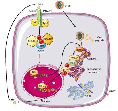 Fig. 1 TAP1's Role in Innate Antiviral Defense.1
Key structural properties of TAP1:
- Pore structure formed by multiple transmembrane α-helices
- Peptide-binding pockets formed by hydrophobic transmembrane regions
- Cytoplasmic side ATP combination box (ABC) structure domain transfer power
Functions of TAP1
The TAP1 gene encodes antigen peptide transporter 1, whose core function is to mediate the transport of endogenous antigen peptides from the cytoplasm to the endoplasmic reticulum, which is a key initiating step in the MHC Class I molecule antigen presentation pathway.
| Function | Description |
| Antigenic peptide transport | Form a heterodimer with TAP2, and use the energy generated by hydrolysis of ATP to actively transport the antigenic peptide generated by proteasome degradation in the cytoplasm to the endoplasmic reticulum cavity. |
| Immune recognition activation | The transported antigenic peptides are assembled with MHC class I molecules in the endoplasmic reticulum and eventually presented to the cell surface for recognition by CD8⁺ T cells, initiating cellular immune responses. |
| Maintenance of immune homeostasis | By continuously presenting autoantigen peptides, it participates in the establishment of central and peripheral tolerance and prevents autoimmune responses. |
| Anti-infection defense | When a virus infects or cells become cancerous, it is responsible for presenting the corresponding abnormal antigen peptides, thereby activating cytotoxic T cells and eliminating diseased cells. |
| Immune editing effect | Its expression level or functional status can affect the antigen presentation efficiency of tumor cells and is an important link in immune editing and tumor immune escape. |
The transport process of TAP1 is substrate selective and ATP-dependent, which is different from the oxygenation balance mechanism of myoglobin. Its functional efficiency directly affects the body's immune surveillance ability against intracellular pathogens and cancer cells.
Applications of TAP1 and TAP1 Antibody in Literature
- Attaran, Nima, et al. "Downregulation of TAP1 in tumor-free tongue contralateral to squamous cell carcinoma of the oral tongue, an indicator of better survival." International journal of molecular sciences 21.17 (2020): 6220. https://doi.org/10.3390/ijms21176220
Research has found that a lower expression of TAP1 mRNA in the contralateral tumor-free tongue tissue of patients with oral tongue squamous cell carcinoma (SCCOT) is significantly associated with better overall survival and disease-free survival, providing a new direction for early diagnosis and prognosis assessment.
- Tabassum, Anika, et al. "Transporter associated with antigen processing 1 (TAP1) expression and prognostic analysis in breast, lung, liver, and ovarian cancer." Journal of Molecular Medicine 99.9 (2021): 1293-1309. https://doi.org/10.1007/s00109-021-02088-w
This study, through bioinformatics analysis, found that the expression, mutation and methylation patterns of the TAP1 gene in different cancers were significantly different, and its expression level was closely related to the prognosis of patients, providing a new basis for understanding the role of TAP1 in tumor progression and chemotherapy resistance.
- Laplana Lafaja, Marina, et al. "Resilience effects of SGK1 and TAP1 DNA markers during PRRSV outbreaks in reproductive sows." International journal of molecular sciences (2020) 10(5), 902. https://doi.org/10.3390/ani10050902
Research has found that specific variations in the SGK1 and TAP1 genes can affect the reproductive performance of sows during an outbreak of porcine reproductive and respiratory syndrome virus (PRRSV). Sows carrying specific genotypes can maintain stable litter sizes and piglet survival rates during the epidemic, providing potential molecular markers for breeding and selection.
- Zhou, Xiao‐Tian, et al. "Hedgehog signalling mediates drug resistance through targeting TAP1 in hepatocellular carcinoma." Journal of Cellular and Molecular Medicine 24.7 (2020): 4298-4311. https://doi.org/10.1111/jcmm.15090
Research has found that the key transcription factor GLI1/2 of the Hedgehog signaling pathway directly regulates the expression of TAP1 and mediates multidrug resistance in liver cancer. Inhibiting this pathway or TAP1 can significantly enhance the sensitivity to chemotherapy drugs such as sorafenib, providing a new target for the treatment of refractory liver cancer.
- Li, Boya, et al. "Antigen peptide transporter 1 (TAP1) promotes resistance to MEK inhibitors in pancreatic cancers." International Journal of Molecular Sciences 23.13 (2022): 7168. https://doi.org/10.3390/ijms23137168
Research has found that antigen peptide transporter 1 (TAP1) leads to drug resistance in pancreatic cancer by promoting the efflux of MEK inhibitors. Inhibiting TAP1 can significantly enhance the sensitivity of tumors to MEK inhibitors, providing a new strategy for combined therapy.
Creative Biolabs: TAP1 Antibodies for Research
Creative Biolabs specializes in the production of high-quality TAP1 antibodies for research and industrial applications. Our portfolio includes monoclonal antibodies tailored for ELISA, Flow Cytometry, Western blot, immunohistochemistry, and other diagnostic methodologies.
- Custom TAP1 Antibody Development: Tailor-made solutions to meet specific research requirements.
- Bulk Production: Large-scale antibody manufacturing for industry partners.
- Technical Support: Expert consultation for protocol optimization and troubleshooting.
- Aliquoting Services: Conveniently sized aliquots for long-term storage and consistent experimental outcomes.
For more details on our TAP1 antibodies, custom preparations, or technical support, contact us at email.
Reference
- Zhao, Jin, et al. "Broadly antiviral activities of TAP1 through activating the TBK1-IRF3-mediated type I interferon production." International Journal of Molecular Sciences 22.9 (2021): 4668. https://doi.org/10.3390/ijms22094668
Anti-TAP1 antibodies
 Loading...


Hot products 
-
Mouse Anti-ALB Recombinant Antibody (V2-55272) (CBMAB-H0819-FY)

-
Mouse Anti-AAV9 Recombinant Antibody (V2-634029) (CBMAB-AP023LY)

-
Mouse Anti-GGT1 Recombinant Antibody (1F9) (CBMAB-G3273-LY)

-
Mouse Anti-FLT1 Recombinant Antibody (11) (CBMAB-V0154-LY)

-
Mouse Anti-ARG1 Recombinant Antibody (CBYCL-103) (CBMAB-L0004-YC)

-
Mouse Anti-CRYAB Recombinant Antibody (A4345) (CBMAB-A4345-YC)

-
Mouse Anti-EMP3 Recombinant Antibody (CBFYE-0100) (CBMAB-E0207-FY)

-
Mouse Anti-BRCA2 Recombinant Antibody (CBYY-0790) (CBMAB-0793-YY)

-
Mouse Anti-CD83 Recombinant Antibody (HB15) (CBMAB-C1765-CQ)

-
Mouse Anti-AKT1 (Phosphorylated S473) Recombinant Antibody (V2-505430) (PTM-CBMAB-0067LY)

-
Mouse Anti-BBS2 Recombinant Antibody (CBYY-0253) (CBMAB-0254-YY)

-
Mouse Anti-ARHGAP5 Recombinant Antibody (54/P190-B) (CBMAB-P0070-YC)

-
Mouse Anti-CTNND1 Recombinant Antibody (CBFYC-2414) (CBMAB-C2487-FY)

-
Mouse Anti-CIITA Recombinant Antibody (CBLC160-LY) (CBMAB-C10987-LY)

-
Mouse Anti-NSUN6 Recombinant Antibody (D-5) (CBMAB-N3674-WJ)

-
Mouse Anti-BCL6 Recombinant Antibody (CBYY-0435) (CBMAB-0437-YY)

-
Mouse Anti-ANXA7 Recombinant Antibody (A-1) (CBMAB-A2941-YC)

-
Mouse Anti-BIRC7 Recombinant Antibody (88C570) (CBMAB-L0261-YJ)

-
Mouse Anti-BIRC3 Recombinant Antibody (315304) (CBMAB-1214-CN)

-
Mouse Anti-DDC Recombinant Antibody (8E8) (CBMAB-0992-YC)

- AActivation
- AGAgonist
- APApoptosis
- BBlocking
- BABioassay
- BIBioimaging
- CImmunohistochemistry-Frozen Sections
- CIChromatin Immunoprecipitation
- CTCytotoxicity
- CSCostimulation
- DDepletion
- DBDot Blot
- EELISA
- ECELISA(Cap)
- EDELISA(Det)
- ESELISpot
- EMElectron Microscopy
- FFlow Cytometry
- FNFunction Assay
- GSGel Supershift
- IInhibition
- IAEnzyme Immunoassay
- ICImmunocytochemistry
- IDImmunodiffusion
- IEImmunoelectrophoresis
- IFImmunofluorescence
- IGImmunochromatography
- IHImmunohistochemistry
- IMImmunomicroscopy
- IOImmunoassay
- IPImmunoprecipitation
- ISIntracellular Staining for Flow Cytometry
- LALuminex Assay
- LFLateral Flow Immunoassay
- MMicroarray
- MCMass Cytometry/CyTOF
- MDMeDIP
- MSElectrophoretic Mobility Shift Assay
- NNeutralization
- PImmunohistologyp-Paraffin Sections
- PAPeptide Array
- PEPeptide ELISA
- PLProximity Ligation Assay
- RRadioimmunoassay
- SStimulation
- SESandwich ELISA
- SHIn situ hybridization
- TCTissue Culture
- WBWestern Blot
