TGFBI Antibodies
Background
The TGFBI gene encodes a secretory extracellular matrix protein, which is mainly expressed in epithelial cells and connective tissues. This protein participates in key biological processes such as maintaining corneal transparency, tissue repair, and cell adhesion and migration by mediating the interaction between cells and the matrix. Research has found that specific mutations in the TGFBI gene are directly related to various corneal dystrophy conditions, such as granular corneal dystrophy and Reis-Bucklers corneal dystrophy. These mutations lead to abnormal protein aggregation, which in turn affects the normal structure and function of the cornea. Since the association between its function and disease was revealed, this gene has become an important model for the study of ophthalmic genetic diseases, deepening people's understanding of the mechanism of action of extracellular matrix proteins in tissue homeostasis and genetic diseases.
Structure of TGFBI
The protein encoded by the TGFBI gene (also known as βig-h3) is a secreted extracellular matrix protein with a molecular weight of approximately 68 kDa. There are certain differences in its molecular weight among different species, mainly due to the balance between the conservation of its core functional domain (such as the FAS1 domain) and species-specific amino acid variations.
| Species | Human | Mouse | Bovine | Rat |
| Molecular Weight (kDa) | About 68.0 | About 67.5 | About 68.2 | About 67.8 |
| Primary Structural Differences | Contains four FAS1 functional domains | Highly homologous sequence, function structure domain conservative | Individual amino acid substitutions are present in the non-domain junction region | The sequence similarity to mouse is extremely high |
This protein is composed of 683 amino acids, and its primary structure contains a signal peptide and four highly conserved FAS1 structural repeat units. Each FAS1 domain (approximately 140 amino acids) forms a unique β -sandwich fold, which serves as the functional basis for its interaction with various extracellular components such as integrins and collagen. These domains are concatenated through flexible linker regions rich in proline, giving the protein as a whole a modular extended conformation rather than a compact spherical structure. Its functional activity depends on the key arginine-glycine-aspartic acid (RGD) motif within a specific domain and the special motif that binds to collagen. These characteristics jointly mediate the regulation of cell adhesion, migration and tissue homeostasis.
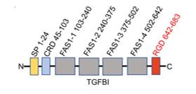 Fig. 1 Schematic diagram of TGFBI protein structure.1
Key structural properties of TGFBI:
- Repetitive sequence of modular FAS1 domains
- Extendable β -folding sandwich structure
- RGD functional motifs with integrin recognition
Functions of TGFBI
The core function of the TGFBI protein (βig-H3) is to mediate cell adhesion, migration and signal transduction in the extracellular matrix. However, it has also been confirmed to be involved in a variety of important pathophysiological processes, including the maintenance of corneal transparency, tissue repair, and tumor development.
| Function | Description |
| Cell adhesion and spreading | The RGD sequence and the FAS1 domain interact with integrins (such as αvβ3 and αvβ5) on the cell surface to mediate cell anchor to the extracellular matrix. |
| Maintenance of corneal transparency | Regular expression and assembly in the subepithelial basement membrane area of the cornea are crucial for maintaining the structural orderliness and optical transparency of the corneal stroma. |
| Tissue damage repair | Expression in the process of wound healing, by promoting fibroblasts and to participate in the formation of cutin cell migration and proliferation tissue remodeling. |
| Regulation of tumor progression | Its effect is environmentally dependent. In some cancers, it can inhibit tumor growth, while in others, it accelerates tumor progression by promoting epithelial-mesenchymal transition and invasion. |
| Hereditary corneal dystrophy | Specific mutations (such as R124H) cause protein misfolding and abnormal aggregation, forming amyloid deposits in the corneal stroma and ultimately damaging vision. |
Unlike myoglobin, which has a clear and single oxygen-binding function, the function of TGFBI protein is highly dependent on the cellular microenvironment in which it is located and the types of receptors it interacts with, showing significant situational dependence and functional diversity. Its dual role in maintaining normal tissue homeostasis and participating in the occurrence of diseases is the focus of current research.
Applications of TGFBI and TGFBI Antibody in Literature
1. Lecker, Laura SM, et al. "TGFBI production by macrophages contributes to an immunosuppressive microenvironment in ovarian cancer." Cancer research 81.22 (2021): 5706-5719. https://doi.org/10.1158/0008-5472.CAN-21-0536
Research has revealed that TGFBI in precancerous lesions of the fallopian tubes is mainly secreted by macrophages, which affect the tumor immune microenvironment through integrin αvβ3 and promote the progression of ovarian cancer. Antibodies targeting TGFBI can inhibit tumor growth in mice, suggesting its potential as a therapeutic target.
2. Chen, Zirong, et al. "Hypoxia-induced TGFBI maintains glioma stem cells by stabilizing EphA2." Theranostics 14.15 (2024): 5778. https://doi.org/10.7150/thno.95141
Research has found that glioma stem cells secrete TGFBI by themselves in a hypoxic environment, activate the AKT-c-MYC pathway by stabilizing the EphA2 protein, and maintain their stem cell characteristics. Targeting the TGFBI/EphA2 axis can inhibit tumor growth, providing a new treatment strategy.
3. Peng, Peng, et al. "TGFBI secreted by tumor-associated macrophages promotes glioblastoma stem cell-driven tumor growth via integrin αvβ5-Src-Stat3 signaling." Theranostics 12.9 (2022): 4221. https://doi.org/10.7150/thno.69605
Studies have confirmed that TGFBI secreted by M2-type macrophages in glioma maintains the characteristics of tumor stem cells and promotes malignant progression by activating the integrin αvβ5-Src-Stat3 pathway. The levels of TGFBI in blood or cerebrospinal fluid can serve as potential diagnostic markers.
4. Lee, Seul Gi, et al. "TGFBI remodels adipose metabolism by regulating the Notch-1 signaling pathway." Experimental & molecular medicine 55.3 (2023): 520-531. https://doi.org/10.1038/s12276-023-00947-9
Research has found that the extracellular matrix protein TGFBI inhibits the "Browning" of adipose tissue and aggravates obesity-related metabolic disorders by activating the Notch-1 pathway in adipocytes. Knockout of TGFBI can improve metabolic health and provide a new target for treatment.
5. Chao-Shern, Connie, et al. "Evaluation of TGFBI corneal dystrophy and molecular diagnostic testing." Eye 33.6 (2019): 874-881. https://doi.org/10.1038/s41433-019-0346-x
Research has found that corneal stromal dystrophy is mainly caused by mutations in the TGFBI gene. Global data analysis shows that existing commercial testing can cover approximately 75% of cases. Research has found that expanding the detection range from 5 hotspot mutations to 11 is expected to increase the detection rate to 90%, which is beneficial to global LASIK preoperative screening.
Creative Biolabs: TGFBI Antibodies for Research
Creative Biolabs specializes in the production of high-quality TGFBI antibodies for research and industrial applications. Our portfolio includes monoclonal antibodies tailored for ELISA, Flow Cytometry, Western blot, immunohistochemistry, and other diagnostic methodologies.
- Custom TGFBI Antibody Development: Tailor-made solutions to meet specific research requirements.
- Bulk Production: Large-scale antibody manufacturing for industry partners.
- Technical Support: Expert consultation for protocol optimization and troubleshooting.
- Aliquoting Services: Conveniently sized aliquots for long-term storage and consistent experimental outcomes.
For more details on our TGFBI antibodies, custom preparations, or technical support, contact us at email.
Reference
- Peng, Peng, et al. "TGFBI secreted by tumor-associated macrophages promotes glioblastoma stem cell-driven tumor growth via integrin αvβ5-Src-Stat3 signaling." Theranostics 12.9 (2022): 4221. https://doi.org/10.7150/thno.69605
Anti-TGFBI antibodies
Products List
 Loading...


Hot products 
-
Mouse Anti-BPGM Recombinant Antibody (CBYY-1806) (CBMAB-2155-YY)

-
Mouse Anti-AAV8 Recombinant Antibody (V2-634028) (CBMAB-AP022LY)

-
Mouse Anti-CD33 Recombinant Antibody (P67.6) (CBMAB-C10189-LY)

-
Mouse Anti-GFAP Recombinant Antibody (5) (CBMAB-G0346-LY)

-
Mouse Anti-AP4E1 Recombinant Antibody (32) (CBMAB-A2996-YC)

-
Rabbit Anti-AKT2 (Phosphorylated S474) Recombinant Antibody (V2-556130) (PTM-CBMAB-0605LY)

-
Mouse Anti-NSUN6 Recombinant Antibody (D-5) (CBMAB-N3674-WJ)

-
Mouse Anti-BIRC5 Recombinant Antibody (6E4) (CBMAB-CP2646-LY)

-
Rat Anti-EMCN Recombinant Antibody (28) (CBMAB-E0280-FY)

-
Mouse Anti-BCL2L1 Recombinant Antibody (H5) (CBMAB-1025CQ)

-
Mouse Anti-AAV9 Recombinant Antibody (V2-634029) (CBMAB-AP023LY)

-
Human Anti-SARS-CoV-2 Spike Recombinant Antibody (CBC05) (CBMAB-CR005LY)

-
Mouse Anti-DISP2 Monoclonal Antibody (F66A4B1) (CBMAB-1112CQ)

-
Mouse Anti-BIRC7 Recombinant Antibody (88C570) (CBMAB-L0261-YJ)

-
Mouse Anti-ACLY Recombinant Antibody (V2-179314) (CBMAB-A0610-YC)

-
Mouse Anti-C4B Recombinant Antibody (CBYY-C2996) (CBMAB-C4439-YY)

-
Mouse Anti-B2M Recombinant Antibody (CBYY-0050) (CBMAB-0050-YY)

-
Mouse Anti-FYN Recombinant Antibody (10) (CBMAB-S6332-CQ)

-
Mouse Anti-DLL4 Recombinant Antibody (D1090) (CBMAB-D1090-YC)

-
Mouse Anti-CD247 Recombinant Antibody (6B10.2) (CBMAB-C1583-YY)

- AActivation
- AGAgonist
- APApoptosis
- BBlocking
- BABioassay
- BIBioimaging
- CImmunohistochemistry-Frozen Sections
- CIChromatin Immunoprecipitation
- CTCytotoxicity
- CSCostimulation
- DDepletion
- DBDot Blot
- EELISA
- ECELISA(Cap)
- EDELISA(Det)
- ESELISpot
- EMElectron Microscopy
- FFlow Cytometry
- FNFunction Assay
- GSGel Supershift
- IInhibition
- IAEnzyme Immunoassay
- ICImmunocytochemistry
- IDImmunodiffusion
- IEImmunoelectrophoresis
- IFImmunofluorescence
- IGImmunochromatography
- IHImmunohistochemistry
- IMImmunomicroscopy
- IOImmunoassay
- IPImmunoprecipitation
- ISIntracellular Staining for Flow Cytometry
- LALuminex Assay
- LFLateral Flow Immunoassay
- MMicroarray
- MCMass Cytometry/CyTOF
- MDMeDIP
- MSElectrophoretic Mobility Shift Assay
- NNeutralization
- PImmunohistologyp-Paraffin Sections
- PAPeptide Array
- PEPeptide ELISA
- PLProximity Ligation Assay
- RRadioimmunoassay
- SStimulation
- SESandwich ELISA
- SHIn situ hybridization
- TCTissue Culture
- WBWestern Blot
