CEACAM1 Antibodies
Background
CEACAM1 is a widely expressed cell surface glycoprotein belonging to the carcinoembryonic antigen family. The protein encoded by this gene plays a significant role in cell adhesion, immune regulation, and signal transduction. Particularly, it is highly expressed in mucosal epithelium, immune cells, and vascular endothelium. Its molecular structure contains an immunoglobulin-like domain, which can mediate homotypic or heterotypic interactions and participate in cell-to-cell recognition and immune checkpoint regulation. CEACAM1 was first identified in 1987. Subsequent studies gradually revealed its dual regulatory functions in tumor immunity, infection response, and metabolic processes. Its complex expression pattern and diverse biological functions make it an important target for immunotherapy and disease mechanism research, providing a key model for understanding cell communication and immune homeostasis.
Structure of CEACAM1
CEACAM1 is a transmembrane protein belonging to the immunoglobulin superfamily. It has multiple subtypes due to alternative splicing, with its molecular weight ranging from 70 to 110 kDa. The difference in molecular weight mainly results from the number of extracellular immunoglobulin-like domains (3-4), the degree of glycosylation, and the different forms of the transmembrane region. The core three-dimensional structure of this protein is composed of multiple immunoglobulin-like domains (IgV and IgC2 types) connected in series, forming an extended extracellular recognition interface. Its secondary structure is mainly based on the characteristic β-sheet barrels within each domain, stabilized by conserved disulfide bonds. The key regulatory structure is located in the intracellular segment, including the immunoreceptor tyrosine inhibitory motif (ITIM), which can specifically recruit phosphatases such as SHP-1 after phosphorylation, thereby conducting inhibitory signals. This structure is the molecular basis for the immune checkpoint function of CEACAM1.
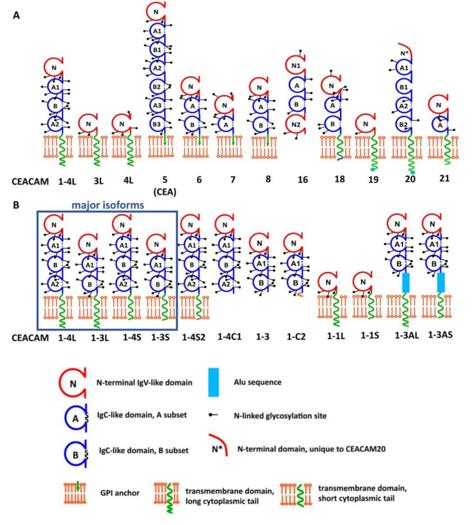 Fig. 1 Structural representation of human CEACAM proteins and CEACAM1 splice variants.1
Key structural properties of CEACAM1:
- Extension of immunoglobulin sample structure domain extracellular area
- Conserved β -folded bucket architectures and in-chain disulfide bonds within each domain
- Cytoplasmic tail contains key immune receptor tyrosine inhibiting motif (ITIM)
Functions of CEACAM1
The core function of CEACAM1 is to act as a multifunctional signal regulator in intercellular communication. Its specific roles are as follows:
| Function | Description |
| Immune Regulation | It transmits inhibitory signals through the cytoplasmic ITIM domain, negatively regulating the activity of immune cells such as T cells and NK cells, and is an important immune checkpoint molecule. |
| Cell Adhesion | It mediates the intercellular recognition and adhesion of the same type (CEACAM1-CEACAM1) or different types (with other members of the CEACAM family), and affects tissue formation and barrier integrity. |
| Vascularization Regulation | Expressed in endothelial cells, it regulates cell proliferation, migration and lumen formation, thereby participating in the balance of vascularization. |
| Pathogen Recognition Receptors | As cell surface receptors for various bacteria (such as Neisseria gonorrhoeae, Helicobacter pylori) and viruses, they mediate the adhesion of pathogens to host cells and the invasion of the host cells. |
| The dual role of tumor suppression and promotion | In normal epithelial tissues, it usually plays a tumor-suppressing role. However, in certain tumor microenvironments, its high expression is instead associated with immune evasion, invasion and metastasis, and its effects are context- and tissue-specific. |
The signal transduction of CEACAM1 depends on the phosphorylation status of its intracellular domain and the interacting partner proteins (such as SHP-1, SHP-2 phosphatases, c-Src kinase, etc.), thereby forming a complex regulatory network that coordinates various physiological and pathological processes such as cell proliferation, differentiation and immune response.
Applications of CEACAM1 and CEACAM1 Antibody in Literature
1. Horst, Andrea Kristina, et al. "CEACAM1 in liver injury, metabolic and immune regulation." International Journal of Molecular Sciences 19.10 (2018): 3110. https://doi.org/10.3390/ijms19103110
The article indicates that CEACAM1 is a transmembrane glycoprotein expressed in epithelial, endothelial and immune cells. It maintains metabolic homeostasis by regulating insulin clearance, and acts as an immune checkpoint to regulate liver inflammation. Its absence can lead to fatty liver, metabolic disorders and exacerbated inflammation, and it plays a central role in liver metabolism and immune balance.
2. Hirao, Hirofumi, et al. "Neutrophil CEACAM1 determines susceptibility to NETosis by regulating the S1PR2/S1PR3 axis in liver transplantation." The Journal of clinical investigation 133.3 (2023). https://doi.org/10.1172/JCI162940
The article indicates that in the middle/small mouse liver transplantation model, the long subtype of neutrophil-specific CEACAM1-L regulates the S1P signaling pathway and autophagy, thereby inhibiting the formation of NETs and alleviating ischemia-reperfusion injury. Clinical data confirm that its expression is negatively correlated with the degree of liver injury and it is a potential immunomodulatory target.
3. Xavier, Serene, et al. "CEACAM1 as a mediator of B-cell receptor signaling in mantle cell lymphoma." Nature Communications 16.1 (2025): 4967. https://doi.org/10.1038/s41467-025-60208-3
This study is the first to reveal that CEACAM1 in mantle cell lymphoma anchors lipid rafts by binding to actin, and recruits SYK kinase to the BCR complex, thereby positively regulating B-cell receptor signal activation. This provides a new idea for targeted therapy.
4. Piloto, Ana Margarida, et al. "Plastic antibodies tailored on quantum dots for an optical detection of myoglobin down to the femtomolar range." Scientific reports 8.1 (2018): 4944. https://doi.org/10.18632/aging.204960
The article indicates that through analysis of the GEO database and experimental verification, it was found that the expression of CEACAM1 was significantly downregulated in oral cancer tissues. Its expression level was closely related to tumor-related pathways and immune infiltration, suggesting that CEACAM1 may play an inhibitory role in the occurrence of oral cancer.
5. Ma, Sai, et al. "CEACAM1 as a molecular target in oral cancer." Aging (Albany NY) 15.16 (2023): 8137. https://doi.org/10.1155/2023/3606362
Based on multi-database analysis, it was found that the expression of CEACAM1 is decreased in renal clear cell carcinoma and is associated with poor prognosis. Its expression level is significantly correlated with tumor progression and the degree of immune cell infiltration, and can serve as a potential prognostic marker and therapeutic target.
Creative Biolabs: CEACAM1 Antibodies for Research
Creative Biolabs specializes in the production of high-quality CEACAM1 antibodies for research and industrial applications. Our portfolio includes monoclonal antibodies tailored for ELISA, Flow Cytometry, Western blot, immunohistochemistry, and other diagnostic methodologies.
- Custom CEACAM1 Antibody Development: Tailor-made solutions to meet specific research requirements.
- Bulk Production: Large-scale antibody manufacturing for industry partners.
- Technical Support: Expert consultation for protocol optimization and troubleshooting.
- Aliquoting Services: Conveniently sized aliquots for long-term storage and consistent experimental outcomes.
For more details on our CEACAM1 antibodies, custom preparations, or technical support, contact us at email.
Reference
- Horst, Andrea Kristina, et al. "CEACAM1 in liver injury, metabolic and immune regulation." International Journal of Molecular Sciences 19.10 (2018): 3110. Distributed under Open Access license CC BY 4.0, without modification. https://doi.org/10.3390/ijms19103110
Anti-CEACAM1 antibodies
 Loading...


Hot products 
-
Mouse Anti-ATP1B1 Recombinant Antibody (E4) (CBMAB-0463-LY)

-
Mouse Anti-ESR1 Recombinant Antibody (Y31) (CBMAB-1208-YC)

-
Mouse Anti-ALOX5 Recombinant Antibody (33) (CBMAB-1890CQ)

-
Mouse Anti-ARID3A Antibody (A4) (CBMAB-0128-YC)

-
Mouse Anti-ACKR3 Recombinant Antibody (V2-261265) (CBMAB-C1023-LY)

-
Mouse Anti-4-Hydroxynonenal Recombinant Antibody (V2-502280) (CBMAB-C1055-CN)

-
Mouse Anti-8-oxoguanine Recombinant Antibody (V2-7697) (CBMAB-1869CQ)

-
Mouse Anti-CARD11 Recombinant Antibody (CBFYC-0811) (CBMAB-C0866-FY)

-
Mouse Anti-Acetyl SMC3 (K105/K106) Recombinant Antibody (V2-634053) (CBMAB-AP052LY)

-
Mouse Anti-ACTN4 Recombinant Antibody (V2-6075) (CBMAB-0020CQ)

-
Mouse Anti-BRCA2 Recombinant Antibody (CBYY-1728) (CBMAB-2077-YY)

-
Mouse Anti-ATP5F1A Recombinant Antibody (51) (CBMAB-A4043-YC)

-
Mouse Anti-CRYAB Recombinant Antibody (A4345) (CBMAB-A4345-YC)

-
Mouse Anti-C5b-9 Recombinant Antibody (aE11) (CBMAB-AO138LY)

-
Mouse Anti-ASB9 Recombinant Antibody (1D8) (CBMAB-A0529-LY)

-
Mouse Anti-AAV-5 Recombinant Antibody (V2-503416) (CBMAB-V208-1402-FY)

-
Mouse Anti-FN1 Monoclonal Antibody (71) (CBMAB-1241CQ)

-
Mouse Anti-DISP2 Monoclonal Antibody (F66A4B1) (CBMAB-1112CQ)

-
Mouse Anti-ALPL Antibody (B4-78) (CBMAB-1009CQ)

-
Mouse Anti-FYN Recombinant Antibody (10) (CBMAB-S6332-CQ)

- AActivation
- AGAgonist
- APApoptosis
- BBlocking
- BABioassay
- BIBioimaging
- CImmunohistochemistry-Frozen Sections
- CIChromatin Immunoprecipitation
- CTCytotoxicity
- CSCostimulation
- DDepletion
- DBDot Blot
- EELISA
- ECELISA(Cap)
- EDELISA(Det)
- ESELISpot
- EMElectron Microscopy
- FFlow Cytometry
- FNFunction Assay
- GSGel Supershift
- IInhibition
- IAEnzyme Immunoassay
- ICImmunocytochemistry
- IDImmunodiffusion
- IEImmunoelectrophoresis
- IFImmunofluorescence
- IGImmunochromatography
- IHImmunohistochemistry
- IMImmunomicroscopy
- IOImmunoassay
- IPImmunoprecipitation
- ISIntracellular Staining for Flow Cytometry
- LALuminex Assay
- LFLateral Flow Immunoassay
- MMicroarray
- MCMass Cytometry/CyTOF
- MDMeDIP
- MSElectrophoretic Mobility Shift Assay
- NNeutralization
- PImmunohistologyp-Paraffin Sections
- PAPeptide Array
- PEPeptide ELISA
- PLProximity Ligation Assay
- RRadioimmunoassay
- SStimulation
- SESandwich ELISA
- SHIn situ hybridization
- TCTissue Culture
- WBWestern Blot
