
CXCR3 Antibodies
Background
CXCR3 is a G protein-coupled receptor mainly expressed in immune cells such as T lymphocytes and natural killer cells, mediating cell migration and immune response by specifically binding to chemokines CXCL9, CXCL10 and CXCL11. This gene plays a crucial role in the regulation of inflammatory responses, autoimmune diseases and the tumor microenvironment, especially in the directional migration of T cells to infected or injured sites. The human CXCR3 gene was first cloned in 1996. Different subtypes produced by its selective splicing, such as CXCR3-A and CXCR3-B, were found to have opposite physiological functions. This characteristic provides an important target for the development of immunomodulatory drugs. In-depth research on the CXCR3 signaling pathway not only reveals the molecular mechanism of immune cell migration but also provides new intervention strategies for the treatment of immune-related diseases such as multiple sclerosis and rheumatoid arthritis.
Structure of CXCR3
Myoglobin is a relatively small protein with a molecular weight of approximately 16.7 kDa. This weight may slightly vary between species due to minor differences in amino acid sequence.
| Species | Human | Mouse | Rat |
| Molecular Weight (kDa) | 40.6 | 40.8 | 40.5 |
| Primary Structural Differences | Splicing variants such as CXCR3-A and CXCR3-B exist | The homology to human is high and the ligand-binding domain is highly conserved | Intracellular area individual amino acid sequence is replaced |
This receptor is composed of 368 amino acids and has a typical seven-time transmembrane domain. Its N-terminal is located outside the cell and contains an important sulfated tyrosine site, which is responsible for specific binding to chemokine ligands such as CXCL9, CXCL10, and CXCL11. The third intracellular loop and the C-terminal region are responsible for coupling G proteins, thereby initiating downstream signal transduction. The ligand-binding pocket of CXCR3 is composed of transmembrane segments, and its activation mechanism depends on the conformational changes of the extracellular loop and the phosphorylation of the key intracellular threonine residues.
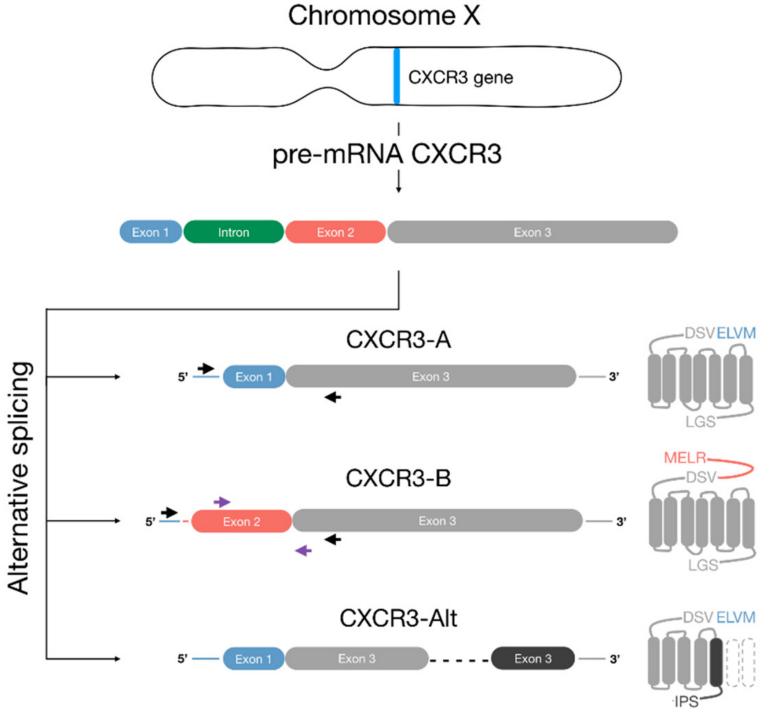 Fig. 1 Schematic representation of CXCR3 variants.1
Key structural properties of CXCR3:
- Typical seven-fold transmembrane (7TM) domain configuration
- Extracellular rich in acidic amino acid N end, used for chemokines specificity recognition
- The third intracellular loop (ICL3) and the C-terminal tail segment are responsible for G protein coupling and signal transduction
Functions of CXCR3
The main function of CXCR3 is to mediate the chemotactic migration and activation of immune cells. However, it is also widely involved in a variety of pathophysiological processes, including inflammatory responses, autoimmune diseases and tumor immune surveillance.
| Function | Description |
| Chemotaxis of immune cells | By combining chemokines such as CXCL9/10/11, activated T cells and NK cells are targeted and recruited to the inflammatory site. |
| Regulation of cell activation | Activate the downstream G protein and Arrestin signaling pathways to regulate the proliferation of lymphocytes, cytokine secretion and effector functions. |
| Inhibition of angiogenesis | The CXCR3-B subtype can inhibit endothelial cell proliferation by binding to a unique ligand, thereby hindering pathological angiogenesis. |
| Inflammation maintenance and regulation | Continuously recruit lymphocytes in chronic inflammation and autoimmune diseases to maintain the formation and persistence of the inflammatory microenvironment. |
| Bidirectional regulation of tumor immunity | Anti-tumor effector cells can be recruited in different tumor types and may also promote tumor metastasis, showing background dependence. |
The signal response of CXCR3 shows G protein preference, which is different from the synergistic effect of hemoglobin. Its ligand binding exhibits high specificity and diversity of signaling pathways, reflecting its precision and functional complexity in immune regulation.
Applications of CXCR3 and CXCR3 Antibody in Literature
1. Karin, Nathan. "CXCR3 ligands in cancer and autoimmunity, chemoattraction of effector T cells, and beyond." Frontiers in immunology 11 (2020): 976. https://doi.org/10.3389/fimmu.2020.00976
The article indicates that CXCR3 mediates the migration and functional differentiation of CD4+ and CD8+T cells through its ligands CXCL9, CXCL10 and CXCL11. CXCL10 enhances the anti-tumor immunity of effector T cells, and CXCL11 promotes the differentiation of regulatory T cells. Both regulate T cell activity through differential bias signal transduction and play a key role in tumors and autoimmunity.
2. Wang, Fangyuan, et al. "The Role of CXCR3 in Nervous System‐Related Diseases." Mediators of Inflammation 2024.1 (2024): 8347647. https://doi.org/10.1155/2024/8347647
The article indicates that CXCR3 and its ligands are widely expressed in the peripheral and central nervous systems and are involved in the occurrence and development of neurological diseases through core pathways such as MAPK. CXCR3 has dual functions of damage and repair in inflammation. Its specific functions vary depending on the cell and disease background, and there is no clear consensus yet.
3. NICLOU, Simone P., Bassam JANJI, and Martyna SZPAKOWSKA. "The Distinct Roles of CXCR3 Variants and Their Ligands in the Tumor Microenvironment." Cells 8.6 (2019). https://doi.org/10.3390/cells8060613
The article indicates that CXCR3 plays A dual role in the tumor microenvironment through its two variants, CXCR3-a and CXCR3-B, along with their corresponding ligands: CXCR3-A can recruit white blood cells to inhibit tumors, while tumors promote progression by altering the expression of CXCR3 and its ligands. This article reviews its tumor suppressor and tumor-promoting mechanisms in tumors.
4. Chan, Travis Yui Hei, et al. "The duality of CXCR3 in glioblastoma: unveiling autocrine and paracrine mechanisms for novel therapeutic approaches." Cell Death & Disease 14.12 (2023): 835. https://doi.org/10.1038/s41419-023-06354-2
The article indicates that CXCR3 and its ligands exert dual functions in glioblastoma (GBM) through autocrine and paracrine effects, not only recruiting and regulating T cells but also participating in tumor progression. Monotherapy and combination therapies targeting the CXCR3 pathway have shown therapeutic potential and are expected to overcome the limitations of current GBM immunotherapy.
5. Szentes, Veronika, et al. "The role of CXCR3 and associated chemokines in the development of atherosclerosis and during myocardial infarction." Frontiers in immunology 9 (2018): 1932. https://doi.org/10.3389/fimmu.2018.01932
The article indicates that CXCR3 and its ligands CXCL9/10/11 play a key role in atherosclerosis and myocardial infarction. By promoting Th1 cell chemotaxis and plaque aggregation, enhancing inflammatory responses, and influencing monocyte/neutrophil recruitment, they are potential therapeutic targets for cardiovascular diseases.
Creative Biolabs: CXCR3 Antibodies for Research
Creative Biolabs specializes in the production of high-quality CXCR3 antibodies for research and industrial applications. Our portfolio includes monoclonal antibodies tailored for ELISA, Flow Cytometry, Western blot, immunohistochemistry, and other diagnostic methodologies.
- Custom CXCR3 Antibody Development: Tailor-made solutions to meet specific research requirements.
- Bulk Production: Large-scale antibody manufacturing for industry partners.
- Technical Support: Expert consultation for protocol optimization and troubleshooting.
- Aliquoting Services: Conveniently sized aliquots for long-term storage and consistent experimental outcomes.
For more details on our CXCR3 antibodies, custom preparations, or technical support, contact us at email.
Reference
- NICLOU, Simone P., Bassam JANJI, and Martyna SZPAKOWSKA. "The Distinct Roles of CXCR3 Variants and Their Ligands in the Tumor Microenvironment." Cells 8.6 (2019). https://doi.org/10.3390/cells8060613
Anti-CXCR3 antibodies
 Loading...
 Compare




Hot products 
-
Mouse Anti-APP Recombinant Antibody (DE2B4) (CBMAB-1122-CN)

-
Mouse Anti-GPRC5C Recombinant Antibody (CBFYH-0464) (CBMAB-H0521-FY)

-
Mouse Anti-ESR1 Recombinant Antibody (Y31) (CBMAB-1208-YC)

-
Mouse Anti-FPR2 Recombinant Antibody (1D6) (CBMAB-F2628-CQ)

-
Mouse Anti-C5AR1 Recombinant Antibody (R63) (CBMAB-C9553-LY)

-
Mouse Anti-AFDN Recombinant Antibody (V2-58751) (CBMAB-L0408-YJ)

-
Mouse Anti-dsRNA Recombinant Antibody (2) (CBMAB-D1807-YC)

-
Mouse Anti-FOXA3 Recombinant Antibody (2A9) (CBMAB-0377-YC)

-
Mouse Anti-C5b-9 Recombinant Antibody (aE11) (CBMAB-AO138LY)

-
Mouse Anti-GNA12 Recombinant Antibody (CBFYH-0402) (CBMAB-H1272-FY)

-
Mouse Anti-GLP1R Recombinant Antibody (4F3) (CBMAB-G0521-LY)

-
Mouse Anti-CORO1A Recombinant Antibody (4G10) (V2LY-1206-LY806)

-
Mouse Anti-APP Recombinant Antibody (5C2A1) (CBMAB-A3314-YC)

-
Mouse Anti-H1-0 Recombinant Antibody (CBFYH-0565) (CBMAB-H1451-FY)

-
Rat Anti-CD63 Recombinant Antibody (7G4.2E8) (CBMAB-C8725-LY)

-
Mouse Anti-ADGRE2 Recombinant Antibody (V2-261270) (CBMAB-C0813-LY)

-
Rat Anti-(1-5)-α-L-Arabinan Recombinant Antibody (V2-501861) (CBMAB-XB0003-YC)

-
Mouse Anti-Gro Monoclonal Antibody (1) (CBMAB-0482-LY)

-
Mouse Anti-CD46 Recombinant Antibody (CBFYC-0076) (CBMAB-C0085-FY)

-
Mouse Anti-ACVR1C Recombinant Antibody (V2-179685) (CBMAB-A1041-YC)

- AActivation
- AGAgonist
- APApoptosis
- BBlocking
- BABioassay
- BIBioimaging
- CImmunohistochemistry-Frozen Sections
- CIChromatin Immunoprecipitation
- CTCytotoxicity
- CSCostimulation
- DDepletion
- DBDot Blot
- EELISA
- ECELISA(Cap)
- EDELISA(Det)
- ESELISpot
- EMElectron Microscopy
- FFlow Cytometry
- FNFunction Assay
- GSGel Supershift
- IInhibition
- IAEnzyme Immunoassay
- ICImmunocytochemistry
- IDImmunodiffusion
- IEImmunoelectrophoresis
- IFImmunofluorescence
- IGImmunochromatography
- IHImmunohistochemistry
- IMImmunomicroscopy
- IOImmunoassay
- IPImmunoprecipitation
- ISIntracellular Staining for Flow Cytometry
- LALuminex Assay
- LFLateral Flow Immunoassay
- MMicroarray
- MCMass Cytometry/CyTOF
- MDMeDIP
- MSElectrophoretic Mobility Shift Assay
- NNeutralization
- PImmunohistologyp-Paraffin Sections
- PAPeptide Array
- PEPeptide ELISA
- PLProximity Ligation Assay
- RRadioimmunoassay
- SStimulation
- SESandwich ELISA
- SHIn situ hybridization
- TCTissue Culture
- WBWestern Blot
