
JAK1 Antibodies
Background
The JAK1 gene encodes a non-receptor tyrosine kinase, which is mainly present in various cells of vertebrates. This protein is involved in cytokine receptor signal transduction and can mediate various physiological processes such as immune regulation, hematopoietic function and inflammatory response. JAK1 plays a key role in autoimmune diseases because the abnormality of its signaling pathway is closely related to various immune system dysfunctions. This gene was first identified in 1990, and the JAK-STAT signaling pathway it mediates is one of the first fully elucidated cytokine signaling mechanisms. Related research led to the recognition of JAK inhibitors for the Nobel Prize in Physiology or Medicine in 2019. Its multi-level regulatory mechanism has become a research hotspot in immunology and tumor treatment, providing an important model for the development of targeted drugs and the understanding of signal networks.
Structure of JAK1
The JAK1 gene encodes a non-receptor tyrosine kinase with a molecular weight of approximately 130 kDa. This protein belongs to the Janus kinase (JAK) family and structurally contains seven homologous domains (JH1-JH7). Among them, the JH1 domain at the carboxyl terminal is a kinase domain responsible for catalyzing phosphorylation reactions, while the adjacent JH2 domain plays a negative regulatory role as a pseudokinase. In signal transduction, the amino-terminal FERM domain (JH4-JH7) of JAK1 binds to the intracellular region of the cytokine receptor. Once the ligand binds to the receptor, JAK1 is activated through mutual phosphorylation, thereby phosphorylating the STAT protein, forming a dimer and translocation into the nucleus to initiate the transcription of the target gene. The three-dimensional structure of this protein presents a typical "bilobar" kinase folding conformation. The conformational changes of the G-loop, αC-helix and activation ring in its active center are crucial for the catalytic function. The kinase domain of JAK1 binds to the pocket of ATP-competitive inhibitors (such as tofatinib) near the hinge region, achieving specific inhibition through hydrogen bond interactions that simulate the adenine ring of ATP.
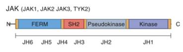 Fig. 1 Homology and functional domain structure of JAKs.1
Key structural properties of JAK1:
- Typical "double leaf" kinase folding structure
- The functional regulatory core consists of the kinase domain (JH1) and pseudokinase domain (JH2)
- The JH1 domain contains a conserved ATP-binding pocket and a catalytic activation ring
Functions of JAK1
The main function of JAK1 is to mediate cytokine signal transduction and regulate immune and hematopoietic functions. In addition, it is also involved in various physiological and pathological processes such as cell proliferation, differentiation and inflammatory responses.
| Function | Description |
| Signal transduction | And cytokine receptor, after the activated protein phosphorylation STAT, start the downstream gene transcription. |
| Immune regulation | Mediating the signaling of multiple key cytokines such as interferons and interleukins is crucial for both innate and adaptive immune responses. |
| Inflammatory regulation | Participate in pro-inflammatory signaling pathways, its excessive activation and rheumatoid arthritis, inflammatory bowel disease such as autoimmune diseases. |
| Hematopoietic support | Play an important role in hematopoietic stem cell proliferation and differentiation, affect the growth of a variety of blood system and function. |
| Tumorigenesis association | The abnormal activation or mutations can lead to continuous proliferation signal, with some blood malignancies and solid tumor development. |
The JAK1-mediated signaling pathway is typically rapid and transient, and its activation depends on the transient binding to the ligand-receptor complex and precise phosphorylation regulation, which contrasts with the pattern of continuous activation of receptor tyrosine kinases, highlighting its core role in the rapid immune response.
Applications of JAK1 and JAK1 Antibody in Literature
1. Sun, Yuechao, Weipeng Gong, and Song Zhang. "METTL3 promotes colorectal cancer progression through activating JAK1/STAT3 signaling pathway." Cell Death & Disease 14.11 (2023): 765. https://doi.org/10.1038/s41419-023-06287-w
The article indicates that METTL3 promotes JAK1 translation through m6A modification and collaborates with NF-κB to activate STAT3 transcription, jointly enhancing the activity of the p-STAT3 pathway and upregulating the expression of VEGFA/CCND1, thereby driving the proliferation and metastasis of colorectal cancer.
2. Pan, Yihui, et al. "EHBP1L1 drives immune evasion in renal cell carcinoma through binding and stabilizing JAK1." Advanced Science 10.11 (2023): 2206792. https://doi.org/10.1002/advs.202206792
The article indicates that EHBP1L1 is highly expressed in renal cell carcinoma, stabilizes JAK1 by competing for SOCS1, activates the JAK1/STAT1/PD-L1 signaling axis, forms an immunosuppressive microenvironment, promotes tumor immune escape and leads to immunotherapy resistance.
3. Nielsen, Ole Haagen, et al. "Selective JAK1 inhibitors for the treatment of inflammatory bowel disease." Pharmacology & therapeutics 245 (2023): 108402. https://doi.org/10.1016/j.pharmthera.2023.108402
The article indicates that JAK inhibitors are used to treat inflammatory bowel disease. Although first-generation drugs such as tofacitinib are effective, they carry cardiovascular risks. The new generation of selective JAK1 inhibitors aims to precisely regulate cytokines, enhance therapeutic efficacy and reduce side effects.
4. Zhang, Huanling, et al. "Endothelial STING-JAK1 interaction promotes tumor vasculature normalization and antitumor immunity." The Journal of Clinical Investigation 135.2 (2025). https://doi.org/10.1172/JCI180622
The article indicates that in endothelial cells, STING mediates the activation of JAK1-STAT signaling through palmitoylation, independent of IFN-γ, promoting vascular normalization and CD8+ T cell infiltration, revealing a new mechanism of anti-tumor immunity by STING agonists.
5. Jensen, Lise Torp, et al. "Allosteric TYK2 inhibition: redefining autoimmune disease therapy beyond JAK1-3 inhibitors." EBioMedicine 97 (2023). https://doi.org/10.1016/j.ebiom.2023.104840
The article indicates that the JAK-STAT pathway has complex functions. Only four JAK kinases mediate 57 cytokine signals, and there is extensive crosstalk. Among them, TYK2 is negatively correlated with various autoimmune diseases due to protective gene variations (such as P1104A), making it a preferred target for drug development.
Creative Biolabs: JAK1 Antibodies for Research
Creative Biolabs specializes in the production of high-quality JAK1 antibodies for research and industrial applications. Our portfolio includes monoclonal antibodies tailored for ELISA, Flow Cytometry, Western blot, immunohistochemistry, and other diagnostic methodologies.
- Custom JAK1 Antibody Development: Tailor-made solutions to meet specific research requirements.
- Bulk Production: Large-scale antibody manufacturing for industry partners.
- Technical Support: Expert consultation for protocol optimization and troubleshooting.
- Aliquoting Services: Conveniently sized aliquots for long-term storage and consistent experimental outcomes.
For more details on our JAK1 antibodies, custom preparations, or technical support, contact us at email.
Reference
- Jensen, Lise Torp, et al. "Allosteric TYK2 inhibition: redefining autoimmune disease therapy beyond JAK1-3 inhibitors." EBioMedicine 97 (2023). https://doi.org/10.1016/j.ebiom.2023.104840
Anti-JAK1 antibodies
 Loading...
 Compare




Hot products 
- AActivation
- AGAgonist
- APApoptosis
- BBlocking
- BABioassay
- BIBioimaging
- CImmunohistochemistry-Frozen Sections
- CIChromatin Immunoprecipitation
- CTCytotoxicity
- CSCostimulation
- DDepletion
- DBDot Blot
- EELISA
- ECELISA(Cap)
- EDELISA(Det)
- ESELISpot
- EMElectron Microscopy
- FFlow Cytometry
- FNFunction Assay
- GSGel Supershift
- IInhibition
- IAEnzyme Immunoassay
- ICImmunocytochemistry
- IDImmunodiffusion
- IEImmunoelectrophoresis
- IFImmunofluorescence
- IGImmunochromatography
- IHImmunohistochemistry
- IMImmunomicroscopy
- IOImmunoassay
- IPImmunoprecipitation
- ISIntracellular Staining for Flow Cytometry
- LALuminex Assay
- LFLateral Flow Immunoassay
- MMicroarray
- MCMass Cytometry/CyTOF
- MDMeDIP
- MSElectrophoretic Mobility Shift Assay
- NNeutralization
- PImmunohistologyp-Paraffin Sections
- PAPeptide Array
- PEPeptide ELISA
- PLProximity Ligation Assay
- RRadioimmunoassay
- SStimulation
- SESandwich ELISA
- SHIn situ hybridization
- TCTissue Culture
- WBWestern Blot
