MAT2A Antibodies
Background
The MAT2A gene encodes methionine adenosine transferase, which acts as a key metabolic enzyme and is mainly present in mammalian tissues such as the liver and kidneys. This enzyme catalyzes the conversion of methionine to S-adenosylmethionine, a reaction that supports the methylation metabolic process within cells and is crucial for DNA modification and signal transduction. The proliferation and differentiation of cells are highly dependent on the methylation donor balance maintained by MAT2A, and this gene often shows abnormal expression in malignant tumors. The crystal structure of this gene was resolved in the 1990s, which facilitated the development of targeted drugs for methylation. The highly conserved structural and functional mechanism of this gene provides an important molecular model for studying epigenetic regulation, metabolic reprogramming, and tumor treatment.
Structure of MAT2A
The methionine adenosine transferase encoded by the MAT2A gene is a relatively small homotrimeric protein, with a monomer molecular weight of approximately 43.7 kDa. The molecular weight of this enzyme is relatively conserved among different species, but there are subtle differences in amino acid sequences.
| Species | Human | Mouse | Rat | Bovine |
| Molecular Weight (kDa) | 43.7 | 43.5 | 43.8 | 43.6 |
| Primary Structural Differences | The catalytic domain is highly conserved | The sequence of active sites is identical | Individual synonymous substitutions exist | Has an extremely high similarity to human enzyme sequences |
This enzyme is composed of approximately 395 amino acids and exhibits a typical α/β folded spherical structure. Its active center contains a crucial methionine-binding pocket, which is composed of multiple conserved amino acid residues (such as aspartic acid and histidine), all of which participate in the catalytic reaction. The quaternary structure of the enzyme forms a functional homologous tetramer, which is the structural basis necessary for its efficient catalysis and the conversion of methionine and ATP into S-adenosylmethionine.
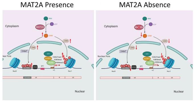 Fig. 1 Schematic diagram showing the role of MAT2A in the regulation of HIV-1 latency.1
Key structural properties of MAT2A:
- Typical α/β folded spherical catalytic domain
- Conservative methionine and ATP dual-substrate binding pocket
- Key aspartic acid residues are involved in the catalytic sulfur transfer reaction
Functions of MAT2A
The main function of the protein encoded by the MAT2A gene is to catalyze the biosynthesis of S-adenosylmethionine (SAM), which is the most important methyl donor in the cell. Additionally, this enzyme is involved in regulating a variety of crucial cellular physiological processes.
| Function | Description |
| Methyl donor synthesis | Catalyzes the reaction of methionine with ATP to form SAM, providing methyl groups for the methylation reactions of DNA, RNA, proteins and phospholipids. |
| Metabolic Regulation | By controlling the level of SAM, it affects the one-carbon metabolism cycle and connects amino acid metabolism with nucleotide synthesis. |
| Cell Proliferation Support | Provide sufficient methyl groups to rapidly dividing cells (such as stem cells and tumor cells), supporting the synthesis and modification of biological macromolecules. |
| Epigenetic Regulation | The product SAM is a necessary substrate for histone and DNA methyltransferases, directly participating in the programming and regulation of gene expression. |
| Liver Function Maintenance | It has extremely high activity in the liver and participates in detoxification processes, bile synthesis, and lipid metabolism through methylation modification. |
The catalytic kinetics of this enzyme exhibits the typical characteristics of a Michaelis enzyme. Its activity is inhibited by the allosteric feedback of the product SAM. This regulatory mechanism ensures the homeostatic balance of methyl donors within the cell, which is fundamentally different from the synergistic allosteric regulation of hemoglobin.
Applications of MAT2A and MAT2A Antibody in Literature
1. Yang, Xiaofan, et al. "MAT2A-mediated S-adenosylmethionine level in CD4+ T cells regulates HIV-1 latent infection." Frontiers in Immunology 12 (2021): 745784. https://doi.org/10.3389/fimmu.2021.745784
This study identified through CRISPR screening that MAT2A regulates one-carbon metabolism to affect the latency of HIV-1. Knockout of MAT2A reduces DNA in the 5'-LTR region and histone methylation, promoting virus activation. Its metabolite SAM is associated with the patient's viral reservoir, providing a new target for functional HIV cure.
2. Chu, Pei-Yi, et al. "MAT2A localization and its independently prognostic relevance in breast cancer patients." International journal of molecular sciences 22.10 (2021): 5382. https://doi.org/10.3390/ijms22105382
The study found that the ratio of cytoplasmic/nuclear expression of MAT2A in breast cancer cells is an independent prognostic indicator. A ratio of ≥ 1.0 indicates a lower survival rate for the patients and is associated with an increased invasiveness of the cancer cells, making it a new prognostic marker.
3. Jiang, Lishan, et al. "MAT2A inhibition suppresses inflammation in Porphyromonas gingivalis-infected human gingival fibroblasts." Journal of Oral Microbiology 16.1 (2024): 2292375. https://doi.org/10.1080/20002297.2023.2292375
The study found that the methionine metabolism mediated by MAT2A promotes the inflammatory response caused by Porphyromonas gingivalis in human gingival fibroblasts. Inhibiting MAT2A can down-regulate the NF-κB/MAPK pathway and reduce the expression of inflammatory factors, providing a new target for the treatment of periodontitis.
4. Xiao, Wanli, et al. "Inhibition of MAT2A impairs skeletal muscle repair function." Biomolecules 14.9 (2024): 1098. https://doi.org/10.3390/biom14091098
The research has found that aging leads to a decrease in the expression of MAT2A in skeletal muscle, which impairs the muscle's regenerative and repair capabilities. The mechanism involves an increase in cell apoptosis mediated by the p53-Fas pathway. Supplementing with its product SAM can improve this situation.
5. Wan, Xinyi, et al. "MAT2B regulates the protein level of MAT2A to preserve RNA N6-methyladenosine." Cell Death & Disease 15.10 (2024): 714. https://doi.org/10.1038/s41419-024-07093-8
The research has found that MAT2B stabilizes MAT2A through NADP+ binding and regulates SAM synthesis and mRNA m6A modification. In liver cancer, inhibiting the interaction between MAT2B and MAT2A can hinder tumor growth, revealing the impact of metabolic interactions on tumor progression.
Creative Biolabs: MAT2A Antibodies for Research
Creative Biolabs specializes in the production of high-quality MAT2A antibodies for research and industrial applications. Our portfolio includes monoclonal antibodies tailored for ELISA, Flow Cytometry, Western blot, immunohistochemistry, and other diagnostic methodologies.
- Custom MAT2A Antibody Development: Tailor-made solutions to meet specific research requirements.
- Bulk Production: Large-scale antibody manufacturing for industry partners.
- Technical Support: Expert consultation for protocol optimization and troubleshooting.
- Aliquoting Services: Conveniently sized aliquots for long-term storage and consistent experimental outcomes.
For more details on our MAT2A antibodies, custom preparations, or technical support, contact us at email.
Reference
- Yang, Xiaofan, et al. "MAT2A-mediated S-adenosylmethionine level in CD4+ T cells regulates HIV-1 latent infection." Frontiers in Immunology 12 (2021): 745784. https://doi.org/10.3389/fimmu.2021.745784
Anti-MAT2A antibodies
 Loading...


Hot products 
-
Mouse Anti-GFAP Recombinant Antibody (5) (CBMAB-G0346-LY)

-
Mouse Anti-ADAM29 Recombinant Antibody (V2-179787) (CBMAB-A1149-YC)

-
Mouse Anti-AKR1B1 Antibody (V2-2449) (CBMAB-1001CQ)

-
Mouse Anti-BIRC3 Recombinant Antibody (315304) (CBMAB-1214-CN)

-
Mouse Anti-BHMT Recombinant Antibody (CBYY-0547) (CBMAB-0550-YY)

-
Rat Anti-CD300A Recombinant Antibody (172224) (CBMAB-C0423-LY)

-
Mouse Anti-B2M Recombinant Antibody (CBYY-0050) (CBMAB-0050-YY)

-
Rabbit Anti-AKT3 Recombinant Antibody (V2-12567) (CBMAB-1057-CN)

-
Mouse Anti-FeLV g27 Recombinant Antibody (1) (CBMAB-V208-1714-FY)

-
Armenian hamster Anti-CD40 Recombinant Antibody (HM40-3) (CBMAB-C10365-LY)

-
Mouse Anti-4-Hydroxynonenal Recombinant Antibody (V2-502280) (CBMAB-C1055-CN)

-
Mouse Anti-ARSA Recombinant Antibody (CBYC-A799) (CBMAB-A3679-YC)

-
Mouse Anti-ATP5F1A Recombinant Antibody (51) (CBMAB-A4043-YC)

-
Rabbit Anti-ABL1 (Phosphorylated Y185) Recombinant Antibody (V2-443434) (PTM-CBMAB-0001YC)

-
Mouse Anti-dsDNA Recombinant Antibody (22) (CBMAB-AP1954LY)

-
Mouse Anti-BIRC3 Recombinant Antibody (16E63) (CBMAB-C3367-LY)

-
Mouse Anti-APP Recombinant Antibody (DE2B4) (CBMAB-1122-CN)

-
Mouse Anti-APCS Recombinant Antibody (CBYC-A663) (CBMAB-A3054-YC)

-
Mouse Anti-GDF5 Recombinant Antibody (1F4) (CBMAB-G2740-LY)

-
Rabbit Anti-AP2M1 (Phosphorylated T156) Recombinant Antibody (D4F3) (PTM-CBMAB-0610LY)

- AActivation
- AGAgonist
- APApoptosis
- BBlocking
- BABioassay
- BIBioimaging
- CImmunohistochemistry-Frozen Sections
- CIChromatin Immunoprecipitation
- CTCytotoxicity
- CSCostimulation
- DDepletion
- DBDot Blot
- EELISA
- ECELISA(Cap)
- EDELISA(Det)
- ESELISpot
- EMElectron Microscopy
- FFlow Cytometry
- FNFunction Assay
- GSGel Supershift
- IInhibition
- IAEnzyme Immunoassay
- ICImmunocytochemistry
- IDImmunodiffusion
- IEImmunoelectrophoresis
- IFImmunofluorescence
- IGImmunochromatography
- IHImmunohistochemistry
- IMImmunomicroscopy
- IOImmunoassay
- IPImmunoprecipitation
- ISIntracellular Staining for Flow Cytometry
- LALuminex Assay
- LFLateral Flow Immunoassay
- MMicroarray
- MCMass Cytometry/CyTOF
- MDMeDIP
- MSElectrophoretic Mobility Shift Assay
- NNeutralization
- PImmunohistologyp-Paraffin Sections
- PAPeptide Array
- PEPeptide ELISA
- PLProximity Ligation Assay
- RRadioimmunoassay
- SStimulation
- SESandwich ELISA
- SHIn situ hybridization
- TCTissue Culture
- WBWestern Blot
