MDFI Antibodies
Background
The MyoD family inhibitory factor protein encoded by the MDFI gene is a transcriptional regulatory protein mainly present in the cell nuclei of vertebrates. This protein inhibits the expression of muscle-specific genes by binding to MyoD and other myogenic regulatory factors, thereby playing a negative regulatory role in the process of muscle differentiation. Studies have shown that MDFI participates in the protein interaction network through its zinc finger domain, helping to maintain the undifferentiated state of muscle precursor cells, and regulating cell fate decisions during muscle regeneration, embryonic development, and other processes. Since its discovery in the 1990s, this gene has received continuous attention due to its "braking" function in the muscle differentiation pathway. The research on its regulatory mechanism has deepened our understanding of the control of cell differentiation timing, the assembly of transcriptional repression complexes, and developmental plasticity.
Structure of MDFI
The MyoD family inhibitory factor protein encoded by the MDFI gene is a nuclear regulatory protein with a molecular weight of approximately 50-55 kDa. The molecular weight varies among different species, mainly due to the changes in its amino acid sequence and domain composition.
| Species | Human | Mouse | Rat | Zebrafish | Chicken |
| Molecular Weight (kDa) | ~55 | ~53 | ~54 | ~48 | ~52 |
| Primary Structural Differences | Containing the I-mfa domain and nuclear export signal | Highly conservative, functionally similar | High homology with humans | Having homologous genes and functional differentiation | Expressed during embryonic development |
The core function of the MDFI protein is achieved through its specific domains. Its primary structure contains a conserved I-mfa (MyoD family inhibitor) domain, which physically inhibits the binding of the myogenic regulatory factors (such as MyoD, Myogenin) to DNA and their transcriptional activation functions by directly binding to the helix-loop-helix (bHLH) domain of these factors. As a result, it inhibits the expression of muscle-specific genes. Additionally, the MDFI protein contains a nuclear export signal that enables it to shuttle between the nucleus and the cytoplasm, dynamically regulating its inhibitory effect. Its overall three-dimensional structure helps form a stable interaction interface, precisely regulating the process of muscle differentiation.
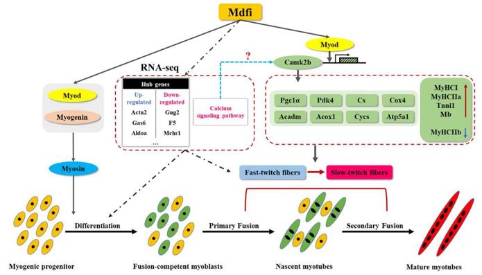 Fig. 1 Mdfi Promotes Myogenic Differentiation and Fast-to-Slow Fiber Switching in C2C12 Cells.1
Key structural properties of MDFI:
- Containing a conserved I-mfa domain (MyoD family inhibitory domain)
- This domain forms an α-helical bundle and can bind to bHLH transcription factors.
- Have approved a signal and output signal, realize the nuclear mass
- Through its hydrophobic interaction interface and MyoD factor, inhibit its transcriptional activity
Functions of MDFI
The main function of the MDFI gene is to act as a transcriptional repressor for muscle differentiation. However, it also plays a role in various physiological processes, including cell proliferation regulation, embryonic development, and tumor formation.
| Function | Description |
| Muscle differentiation inhibition | By directly binding to MyoD and other myogenic regulatory factors through its I-mfa domain, it physically inhibits their transcriptional activity, thereby suppressing the expression of muscle-specific genes. |
| Cell fate maintenance | Highly expressed in muscle precursor cells, it helps maintain the undifferentiated state of the cells and prevents premature differentiation. |
| Embryo Development Regulation | Participates in regulating the formation patterns of various tissues (such as the neural tube and body segments) during the embryonic development process. |
| Tumor Association | In certain cancers (such as rhabdomyosarcoma), it is expressed abnormally, which may affect tumor progression by interfering with the normal differentiation pathway. |
| Signal Pathway Integration | It can interact with components of signaling pathways such as Wnt/β-catenin and play a regulatory role at the intersection of multiple developmental pathways. |
Unlike the linear activation pathways formed by positive regulatory factors such as MyoD, the mechanism of MDFI is more akin to a precise "molecular brake". It achieves efficient and specific inhibition of the initiation of the differentiation process by forming inactive heterodimers or masking DNA binding sites, ensuring the controllability of the temporal sequence of muscle differentiation.
Applications of MDFI and MDFI Antibody in Literature
1. Huang, Bo, et al. "Mdfi promotes C2C12 cell differentiation and positively modulates fast-to-slow-twitch muscle fiber transformation." Frontiers in cell and developmental biology 9 (2021): 605875. https://doi.org/10.3389/fcell.2021.605875
The article indicates that Mdfi promotes myogenic differentiation of C2C12 cells through overexpression of CRISPR/Cas9, and regulates the transformation of fast and slow muscle fibers. Its mechanism of action involves calcium signaling pathways and key genes such as Myod and Camk2b, providing potential targets for the treatment of muscle and metabolic diseases.
2. Chen, Pengyu, et al. "MDFI is a novel biomarker for poor prognosis in LUAD." Frontiers in Oncology 12 (2022): 1005962. https://doi.org/10.3389/fonc.2022.1005962
The study found that MDFI is overexpressed in lung adenocarcinoma (LUAD) tissues and is significantly associated with poor prognosis. MDFI can serve as an independent predictor, and is related to tumor mutation burden and immune infiltration, and is expected to become a potential new biomarker for the diagnosis and prognosis assessment of LUAD.
3. Ma, Ding, et al. "MDFI promotes the proliferation and tolerance to chemotherapy of colorectal cancer cells by binding ITGB4/LAMB3 to activate the AKT signaling pathway." Cancer Biology & Therapy 25.1 (2024): 2314324. https://doi.org/10.1080/15384047.2024.2314324
This study reveals that MDFI, in colorectal cancer, activates the AKT pathway by binding to ITGB4/LAMB3, promoting cancer cell proliferation and reducing their sensitivity to oxaliplatin/fluorouracil chemotherapy. This finding provides a potential molecular target for the treatment of colorectal cancer.
4. Sui, Yuan, et al. "Opposite roles of the JMJD1A interaction partners MDFI and MDFIC in colorectal cancer." Scientific Reports 10.1 (2020): 8710. https://doi.org/10.1038/s41598-020-65536-6
Studies have shown that in colorectal cancer, high expression of MDFI promotes cancer cell proliferation, while low expression of MDFIC inhibits tumor growth. Both interact with JMJD1A. MDFIC exerts an inhibitory function by activating the tumor suppressor gene HIC1, but the expression patterns differ in other cancers.
5. Yanjun, Sun, et al. "The role of miR‐128 and MDFI in cardiac hypertrophy and heart failure: Mechanistic." Journal of Cellular and Molecular Medicine 28.14 (2024): e18546. https://doi.org/10.1111/jcmm.18546
The study found that miR-128 targets and inhibits the expression of MDFI, activating the Wnt1/β-catenin pathway, thereby promoting cardiomyocyte apoptosis and inhibiting their proliferation, and exacerbating heart failure. Inhibiting miR-128 or upregulating MDFI could become potential therapeutic strategies for heart failure.
Creative Biolabs: MDFI Antibodies for Research
Creative Biolabs specializes in the production of high-quality MDFI antibodies for research and industrial applications. Our portfolio includes monoclonal antibodies tailored for ELISA, Flow Cytometry, Western blot, immunohistochemistry, and other diagnostic methodologies.
- Custom MDFI Antibody Development: Tailor-made solutions to meet specific research requirements.
- Bulk Production: Large-scale antibody manufacturing for industry partners.
- Technical Support: Expert consultation for protocol optimization and troubleshooting.
- Aliquoting Services: Conveniently sized aliquots for long-term storage and consistent experimental outcomes.
For more details on our MDFI antibodies, custom preparations, or technical support, contact us at email.
Reference
- Huang, Bo, et al. "Mdfi promotes C2C12 cell differentiation and positively modulates fast-to-slow-twitch muscle fiber transformation." Frontiers in cell and developmental biology 9 (2021): 605875. Distributed under Open Access license CC BY 4.0, without modification. https://doi.org/10.3389/fcell.2021.605875
Anti-MDFI antibodies
 Loading...


Hot products 
-
Mouse Anti-CD24 Recombinant Antibody (HIS50) (CBMAB-C10123-LY)

-
Mouse Anti-ARID1B Recombinant Antibody (KMN1) (CBMAB-A3546-YC)

-
Mouse Anti-CD63 Recombinant Antibody (CBXC-1200) (CBMAB-C1467-CQ)

-
Mouse Anti-AK4 Recombinant Antibody (V2-180419) (CBMAB-A1891-YC)

-
Mouse Anti-APOH Recombinant Antibody (4D9A4) (CBMAB-A3249-YC)

-
Mouse Anti-GLP1R Recombinant Antibody (4F3) (CBMAB-G0521-LY)

-
Mouse Anti-AKR1B1 Antibody (V2-2449) (CBMAB-1001CQ)

-
Rabbit Anti-CAMK2A Recombinant Antibody (BA0032) (CBMAB-0137CQ)

-
Mouse Anti-CD247 Recombinant Antibody (6B10.2) (CBMAB-C1583-YY)

-
Mouse Anti-CARD11 Recombinant Antibody (CBFYC-0811) (CBMAB-C0866-FY)

-
Mouse Anti-FPR2 Recombinant Antibody (1D6) (CBMAB-F2628-CQ)

-
Mouse Anti-ENO1 Recombinant Antibody (CBYC-A950) (CBMAB-A4388-YC)

-
Mouse Anti-CTCF Recombinant Antibody (CBFYC-2371) (CBMAB-C2443-FY)

-
Mouse Anti-ATP1B3 Recombinant Antibody (1E9) (CBMAB-A4021-YC)

-
Mouse Anti-ALB Recombinant Antibody (V2-363290) (CBMAB-S0173-CQ)

-
Mouse Anti-ADRB2 Recombinant Antibody (V2-180026) (CBMAB-A1420-YC)

-
Mouse Anti-ACTG1 Recombinant Antibody (V2-179597) (CBMAB-A0916-YC)

-
Human Anti-SARS-CoV-2 S1 Monoclonal Antibody (CBFYR-0120) (CBMAB-R0120-FY)

-
Mouse Anti-APCS Recombinant Antibody (CBYC-A663) (CBMAB-A3054-YC)

-
Mouse Anti-ASTN1 Recombinant Antibody (H-9) (CBMAB-1154-CN)

- AActivation
- AGAgonist
- APApoptosis
- BBlocking
- BABioassay
- BIBioimaging
- CImmunohistochemistry-Frozen Sections
- CIChromatin Immunoprecipitation
- CTCytotoxicity
- CSCostimulation
- DDepletion
- DBDot Blot
- EELISA
- ECELISA(Cap)
- EDELISA(Det)
- ESELISpot
- EMElectron Microscopy
- FFlow Cytometry
- FNFunction Assay
- GSGel Supershift
- IInhibition
- IAEnzyme Immunoassay
- ICImmunocytochemistry
- IDImmunodiffusion
- IEImmunoelectrophoresis
- IFImmunofluorescence
- IGImmunochromatography
- IHImmunohistochemistry
- IMImmunomicroscopy
- IOImmunoassay
- IPImmunoprecipitation
- ISIntracellular Staining for Flow Cytometry
- LALuminex Assay
- LFLateral Flow Immunoassay
- MMicroarray
- MCMass Cytometry/CyTOF
- MDMeDIP
- MSElectrophoretic Mobility Shift Assay
- NNeutralization
- PImmunohistologyp-Paraffin Sections
- PAPeptide Array
- PEPeptide ELISA
- PLProximity Ligation Assay
- RRadioimmunoassay
- SStimulation
- SESandwich ELISA
- SHIn situ hybridization
- TCTissue Culture
- WBWestern Blot
