PDCD1 Antibodies
Background
The PDCD1 gene encodes a transmembrane glycoprotein called programmed cell death protein 1, which is mainly expressed on the surface of activated T cells, B cells and myeloid cells. This protein regulates the intensity and duration of immune responses by binding to its ligands PD-L1/PD-L2 and transmitting inhibitory signals, thereby maintaining immune tolerance and preventing excessive immune responses. In the tumor microenvironment, tumor cells often highly express PD-L1 to activate the PD-1/PD-L1 pathway, leading to T cell exhaustion. This mechanism makes PD-1 an important target for cancer immunotherapy. This gene was first identified by the team of Japanese scholar Tasuku Honjo in 1992. Its functional research not only revealed the core role of immune checkpoints but also promoted the breakthrough application of monoclonal antibody drugs in the field of tumor treatment. The related achievements won the Nobel Prize in Physiology or Medicine in 2018.
Structure of PDCD1
PDCD1 is an immune checkpoint transmembrane protein with a molecular weight of approximately 55-60 kDa. This value shows slight differences among different species, mainly due to the degree of glycosylation modification and the minor changes in the extracellular immunoglobulin domain (IgV).
| Species | Human | Mouse | Rat | Crab-eating macaque | Dog |
| Molecular Weight (kDa) | 55.4 | 55.1 | 54.8 | 55.6 | 55.2 |
| Primary Structural Differences | Containing 288 amino acids, extracellular area 1 IgV domain structure | Extracellular region has 62% homology with human | Sequence is highly conserved across the membrane area | 92% homology with human PD-1 | Species specific epitopes exist in the IgV domain |
This protein is composed of 288 amino acids, and its extracellular region contains an immunoglobulin-mutable (IgV) domain, which forms a specific spatial conformation through intramolecular disulfide bonds. The intracellular segment carries two characteristic signaling motifs: the tyrosin-based immunosuppressive receptor motif (ITIM) and the tyrosin-based conversion motif (ITSM). These two motifs deliver inhibitory signals by recruiting SHP-2 phosphatase. The entire molecule specifically binds to the ligand PD-L1/PD-L2 through its IgV domain, and this interaction constitutes the structural basis of immune checkpoint blockade therapy.
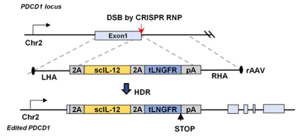
 Fig. 1 Schematic of IL-12 Knock-in at the PDCD1 Locus via CRISPR/AAV6.1
Key structural properties of PDCD1:
- Extracellular immunoglobulin variable region (IgV) domains
- Transmembrane region and intracellular segment containing immunoreceptor inhibitory motif (ITIM) and switching motif (ITSM)
- Cysteine residues form intramolecular disulfide bonds to stabilize the steric conformation
Functions of PDCD1
The main function of PDCD1 (programmed death receptor 1) is to transmit immunosuppressive signals to maintain autoimmune tolerance, while mediating immune escape in the tumor microenvironment.
| Function | Description |
| Regulation of immune homeostasis | SHP-2 phosphatase was recruited through the intracellular ITSM motif to inhibit the activation of the T cell receptor signaling pathway. |
| Maintenance of peripheral tolerance | Limit the excessive activation of effector T cells in lymph nodes and peripheral tissues to prevent autoimmune diseases. |
| T cell exhaustion induction | Continuous expression in chronic infection and tumor environments leads to T cell failure and decreased proliferation capacity. |
| Immune checkpoint function | With PD on the surface of the tumor cells - L1 / L2, help tumor inhibitory signals escape immune clearance. |
| Immunotherapy targets | Monoclonal antibodies restore the anti-tumor activity of T cells by blocking the PD-1/PD-L1 interaction. |
The inhibitory signal intensity of PD-1 is significantly stronger than that of other immune checkpoints such as CTLA-4. Its activation can lead to a significant downregulation of the secretion of key cytokines such as IL-2 and IFN-γ. This characteristic makes it one of the most effective targets in tumor immunotherapy.
Applications of PDCD1 and PDCD1 Antibody in Literature
1. Alexey, Palamarchuk, et al. "PDCD1 (PD-1) is a direct target of miR-15a-5p and miR-16-5p." Signal Transduction and Targeted Therapy 7.1 (2022). https://doi.org/10.1038/s41392-021-00832-9
The article indicates that the PD-1 protein encoded by the PDCD1 gene is a key regulatory receptor for T-cell immunity. Its binding to the ligand PD-L1 can inhibit T cell activity, and various tumors achieve immune escape through this pathway. Antibody drugs targeting PD-1/PD-L1 have achieved remarkable efficacy in the treatment of various cancers.
2. Wang, Yucheng, et al. "Identification of PDCD1 as a potential biomarker in acute rejection after kidney transplantation via comprehensive bioinformatic analysis." Frontiers in Immunology 13 (2023): 1076546. https://doi.org/10.3389/fimmu.2022.1076546
The article indicates that through bioinformatics analysis, it was found that PDCD1 is a key gene for acute kidney transplant rejection. This study revealed that rejection is closely related to T-cell exhaustion and confirmed that PDCD1 can serve as a potential biomarker for diagnosing acute rejection.
3. Goltz, Diane, et al. "PDCD1 (PD-1) promoter methylation predicts outcome in head and neck squamous cell carcinoma patients." Oncotarget 8.25 (2017): 41011. https://doi.org/10.18632/oncotarget.17354
Research has found that hypermethylation of the PDCD1 gene is an independent adverse prognostic factor for head and neck squamous cell carcinoma and is significantly associated with a shortened overall survival period of patients. This indicator may help identify people with a poor prognosis and guide them to receive stronger chemotherapy or immunotherapy.
4. Kobayashi, Mizuki, et al. "Severe immune-related adverse events in patients treated with nivolumab for metastatic renal cell carcinoma are associated with PDCD1 polymorphism." Genes 13.7 (2022): 1204. https://doi.org/10.3390/genes13071204
This study explores the relationship between PDCD1 gene polymorphism and the efficacy of nivolumab in patients with metastatic renal cell carcinoma. The results showed that patients carrying the PD-1.6G allele had a significantly higher risk of serious and multiple immune-related adverse events.
5. Silva, Mauro César da, et al. "Increased PD-1 level in severe cervical injury is associated with the rare programmed cell death 1 (PDCD1) rs36084323 a allele in a dominant model." Frontiers in Cellular and Infection Microbiology 11 (2021): 587932. https://doi.org/10.3389/fcimb.2021.587932
This study explores the role of PDCD1 in HPV-related cervical lesions. Studies have found that the expression of PD-1 protein increases in the area of cervical injury. The -606G>A polymorphism of the PDCD1 gene affects its expression. Patients carrying the A allele have a higher PD-1 level, which may promote the immune escape of HPV.
Creative Biolabs: PDCD1 Antibodies for Research
Creative Biolabs specializes in the production of high-quality PDCD1 antibodies for research and industrial applications. Our portfolio includes monoclonal antibodies tailored for ELISA, Flow Cytometry, Western blot, immunohistochemistry, and other diagnostic methodologies.
- Custom PDCD1 Antibody Development: Tailor-made solutions to meet specific research requirements.
- Bulk Production: Large-scale antibody manufacturing for industry partners.
- Technical Support: Expert consultation for protocol optimization and troubleshooting.
- Aliquoting Services: Conveniently sized aliquots for long-term storage and consistent experimental outcomes.
For more details on our PDCD1 antibodies, custom preparations, or technical support, contact us at email.
Reference
- Kim, Segi, et al. "Reprogramming of IL-12 secretion in the PDCD1 locus improves the anti-tumor activity of NY-ESO-1 TCR-T cells." Frontiers in immunology 14 (2023): 1062365. https://doi.org/10.3389/fimmu.2023.1062365
Anti-PDCD1 antibodies
 Loading...
Hot products 
-
Mouse Anti-CHRNA9 Recombinant Antibody (8E4) (CBMAB-C9161-LY)

-
Mouse Anti-COL1A2 Recombinant Antibody (CF108) (V2LY-1206-LY626)

-
Mouse Anti-BSN Recombinant Antibody (219E1) (CBMAB-1228-CN)

-
Mouse Anti-ACTN4 Recombinant Antibody (V2-6075) (CBMAB-0020CQ)

-
Mouse Anti-AQP2 Recombinant Antibody (G-3) (CBMAB-A3359-YC)

-
Mouse Anti-CTNND1 Recombinant Antibody (CBFYC-2414) (CBMAB-C2487-FY)

-
Mouse Anti-CCNH Recombinant Antibody (CBFYC-1054) (CBMAB-C1111-FY)

-
Mouse Anti-BACE1 Recombinant Antibody (CBLNB-121) (CBMAB-1180-CN)

-
Mouse Anti-DLC1 Recombinant Antibody (D1009) (CBMAB-D1009-YC)

-
Mouse Anti-DMPK Recombinant Antibody (CBYCD-324) (CBMAB-D1200-YC)

-
Rabbit Anti-ADRA1A Recombinant Antibody (V2-12532) (CBMAB-1022-CN)

-
Mouse Anti-AKR1C3 Recombinant Antibody (V2-12560) (CBMAB-1050-CN)

-
Mouse Anti-FN1 Monoclonal Antibody (D6) (CBMAB-1240CQ)

-
Mouse Anti-ACTB Recombinant Antibody (V2-179553) (CBMAB-A0870-YC)

-
Mouse Anti-FLT1 Recombinant Antibody (11) (CBMAB-V0154-LY)

-
Mouse Anti-AKT1/AKT2/AKT3 (Phosphorylated T308, T309, T305) Recombinant Antibody (V2-443454) (PTM-CBMAB-0030YC)

-
Rat Anti-ADAM10 Recombinant Antibody (V2-179741) (CBMAB-A1103-YC)

-
Mouse Anti-BIRC3 Recombinant Antibody (315304) (CBMAB-1214-CN)

-
Mouse Anti-8-oxoguanine Recombinant Antibody (V2-7719) (CBMAB-1898CQ)

-
Mouse Anti-AMACR Recombinant Antibody (CB34A) (CBMAB-CA034LY)

- AActivation
- AGAgonist
- APApoptosis
- BBlocking
- BABioassay
- BIBioimaging
- CImmunohistochemistry-Frozen Sections
- CIChromatin Immunoprecipitation
- CTCytotoxicity
- CSCostimulation
- DDepletion
- DBDot Blot
- EELISA
- ECELISA(Cap)
- EDELISA(Det)
- ESELISpot
- EMElectron Microscopy
- FFlow Cytometry
- FNFunction Assay
- GSGel Supershift
- IInhibition
- IAEnzyme Immunoassay
- ICImmunocytochemistry
- IDImmunodiffusion
- IEImmunoelectrophoresis
- IFImmunofluorescence
- IGImmunochromatography
- IHImmunohistochemistry
- IMImmunomicroscopy
- IOImmunoassay
- IPImmunoprecipitation
- ISIntracellular Staining for Flow Cytometry
- LALuminex Assay
- LFLateral Flow Immunoassay
- MMicroarray
- MCMass Cytometry/CyTOF
- MDMeDIP
- MSElectrophoretic Mobility Shift Assay
- NNeutralization
- PImmunohistologyp-Paraffin Sections
- PAPeptide Array
- PEPeptide ELISA
- PLProximity Ligation Assay
- RRadioimmunoassay
- SStimulation
- SESandwich ELISA
- SHIn situ hybridization
- TCTissue Culture
- WBWestern Blot


