
ENO1 Antibodies
Background
The α-enolase encoded by the ENO1 gene is a multifunctional glycolytic enzyme that is widely present in the cytoplasm of various biological cells. It not only catalyzes the conversion of 2-phosphoglyceric acid to phosphoenolpyruvic acid, but also acts as a fibrinogen receptor on the cell surface to participate in tissue remodeling. This gene also plays a structural protein role in the nervous system, maintaining the transparency of crystals. The abnormal expression of ENO1 is closely related to the occurrence of various tumors, and it enhances the invasive ability of cancer cells by promoting the Warburg effect. The study of ENO1 not only reveals the non-classical functions of metabolic enzymes, but also provides an important basis for the development of cancer diagnostic markers and potential therapeutic targets.
Structure of ENO1
The α-enolase protein encoded by the ENO1 gene has a molecular weight of approximately 47 kDa. There is a slight fluctuation due to sequence differences among different species. This protein consists of 433 amino acids and forms a tight barrel-like structure. Its active center contains a necessary magnesium ion binding site. ENO1 exists in the cytoplasm in a dimeric form, with each subunit containing two domains: a smaller N-terminal domain and a larger C-terminal catalytic domain. During the dehydration of 2-phosphoglyceric acid to form phosphoenolpyruvic acid, two conserved lysine residues participate in proton transfer. In addition to the enzymatic function, the C-terminal region of ENO1 also participates in the interaction with other proteins, mediating its function as a fibrinogen receptor on the cell surface. This protein is upregulated in various tumors. Its post-translational modifications include phosphorylation and acetylation, which can regulate enzyme activity and subcellular localization.
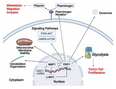 Fig. 1 Schematic representation of ENO1's multiple cellular functions.1
Key structural properties of ENO1:
- Tight barrel structure, consisting of two structural domains
- The active center contains a magnesium ion binding site
- Two pairs of conserved amino acid residues are involved in proton transfer
- Dimeric forms exist, and inter-subunits are stabilized by hydrophobic interactions
- The C-terminal region mediates interactions with other proteins
Functions of ENO1
The α-enolase encoded by the ENO1 gene was initially regarded as a classic glycolytic enzyme, but subsequent studies have revealed that it is involved in various cellular processes:
| Function | Description |
| Glycolysis catalysis | Converts 2-phosphoglyceric acid to phosphoenolpyruvic acid in the cytoplasm, generating ATP for the cell. |
| Low-oxygen adaptation | Expressed up-regulated under hypoxic conditions, binds to specific mRNA to enhance its stability, regulating energy metabolism. |
| Fibrinogen receptor | Located on the cell surface, binds fibrinogen and promotes fibrinolysis, participating in cell migration and tissue remodeling. |
| Self-antigen | Recognized by the immune system in autoimmune diseases, involved in the pathogenesis of diseases such as rheumatoid arthritis. |
| Tumor-related functions | Highly expressed in various cancer cells, enhances tumor growth and metastasis by promoting the Warburg effect. |
| Transcription regulation | Partially located in the cell nucleus, acts as a transcriptional co-activator to participate in gene expression regulation. |
As a multifunctional protein, ENO1 exhibits different functions depending on its cellular localization and post-translational modification status.
Applications of ENO1 and ENO1 Antibody in Literature
1. Huang, Chen Kai, et al. "ENO1 and cancer." Molecular Therapy-Oncolytics 24 (2022): 288-298. https://doi.org/10.1016/j.omto.2021.12.026
The article indicates that ENO1 is a multifunctional glycolytic enzyme that is highly expressed on the surface and in the cytoplasm of cancer cells. It is involved in processes such as tumor proliferation, apoptosis inhibition, immune evasion, and promotes angiogenesis and metastasis. It is a promising tumor marker and an immunotherapy target.
2. Chen, Baoxiang, et al. "Metabolic Recoding of NSUN2‐mediated m5C modification promotes the progression of colorectal cancer via the NSUN2/YBX1/m5C‐ENO1 positive feedback loop." Advanced Science 11.28 (2024): 2309840. https://doi.org/10.1002/advs.202309840
The article indicates that in colorectal cancer, the m5C methyltransferase NSUN2 is highly expressed. By "writing" onto the ENO1 mRNA and recruiting the "reader" YBX1, it promotes glycolysis. The produced lactic acid then feedback-activates NSUN2, forming a positive feedback loop, connecting metabolism with epigenetic regulation, providing new ideas for combined treatment.
3. Zhao, Yun, et al. "CircFndc3b mediates exercise‐induced neuroprotection by mitigating microglial/macrophage pyroptosis via the ENO1/KLF2 axis in stroke mice." Advanced Science 12.1 (2025): 2403818. https://doi.org/10.1002/advs.202403818
The article indicates that exercise induces the increase of circFndc3b in macrophages, which binds to ENO1, enhances the stability of Klf2 and FUS mRNA, inhibits cell pyroptosis, and alleviates cerebral ischemic injury. The study reveals a new mechanism and therapeutic target of ENO1-mediated neuroprotection.
4. Shen, Dexin, et al. "Melatonin inhibits bladder tumorigenesis by suppressing PPARγ/ENO1-mediated glycolysis." Cell Death & Disease 14.4 (2023): 246. https://doi.org/10.1038/s41419-023-05770-8
The article indicates that melatonin downregulates ENO1 expression through PPARγ, inhibits glycolysis in bladder cancer, and enhances the sensitivity to gemcitabine. Its effect is mediated by ROS accumulation, providing a new strategy for overcoming chemotherapy resistance.
5. He, Xueling, et al. "Identifying ENO1 as a protein target of chlorogenic acid to inhibit cellular senescence and prevent skin photoaging in mice." Aging Cell 24.4 (2025): e14433. https://doi.org/10.1111/acel.14433
The article indicates that chlorogenic acid covalently binds to ENO1 to inhibit glycolysis, thereby preventing fibroblast senescence induced by UVA. In a mouse model, it was verified that it can alleviate skin photoaging, suggesting that ENO1 is a new target for anti-photoaging.
Creative Biolabs: ENO1 Antibodies for Research
Creative Biolabs specializes in the production of high-quality ENO1 antibodies for research and industrial applications. Our portfolio includes monoclonal and polyclonal antibodies tailored for ELISA, Flow Cytometry, Western blot, immunohistochemistry, and other diagnostic methodologies.
- Custom ENO1 Antibody Development: Tailor-made solutions to meet specific research requirements.
- Bulk Production: Large-scale antibody manufacturing for industry partners.
- Technical Support: Expert consultation for protocol optimization and troubleshooting.
- Aliquoting Services: Conveniently sized aliquots for long-term storage and consistent experimental outcomes.
For more details on our ENO1 antibodies, custom preparations, or technical support, contact us at email.
Reference
- Huang, Chen Kai, et al. "ENO1 and cancer." Molecular Therapy-Oncolytics 24 (2022): 288-298. Distributed under Open Access license CC BY 4.0, without modification. https://doi.org/10.1016/j.omto.2021.12.026
Anti-ENO1 antibodies
 Loading...
 Compare




Hot products 
- AActivation
- AGAgonist
- APApoptosis
- BBlocking
- BABioassay
- BIBioimaging
- CImmunohistochemistry-Frozen Sections
- CIChromatin Immunoprecipitation
- CTCytotoxicity
- CSCostimulation
- DDepletion
- DBDot Blot
- EELISA
- ECELISA(Cap)
- EDELISA(Det)
- ESELISpot
- EMElectron Microscopy
- FFlow Cytometry
- FNFunction Assay
- GSGel Supershift
- IInhibition
- IAEnzyme Immunoassay
- ICImmunocytochemistry
- IDImmunodiffusion
- IEImmunoelectrophoresis
- IFImmunofluorescence
- IGImmunochromatography
- IHImmunohistochemistry
- IMImmunomicroscopy
- IOImmunoassay
- IPImmunoprecipitation
- ISIntracellular Staining for Flow Cytometry
- LALuminex Assay
- LFLateral Flow Immunoassay
- MMicroarray
- MCMass Cytometry/CyTOF
- MDMeDIP
- MSElectrophoretic Mobility Shift Assay
- NNeutralization
- PImmunohistologyp-Paraffin Sections
- PAPeptide Array
- PEPeptide ELISA
- PLProximity Ligation Assay
- RRadioimmunoassay
- SStimulation
- SESandwich ELISA
- SHIn situ hybridization
- TCTissue Culture
- WBWestern Blot
