PRKDC Antibodies
Background
The PRKDC gene encodes the catalytic subunit of DNA-dependent protein kinase, which is a large protein kinase that plays a central role in DNA double-strand break repair. This protein, as the catalytic subunit of the DNA-PK complex, is activated by the Ku70/Ku80 heterodimer upon recognition of DNA damage and initiates the non-homologous end joining repair pathway. It is also a key factor in the V(D)J recombination of the immune system. Mutations in the PRKDC gene can lead to severe combined immunodeficiency, characterized by the retardation of T-cell and B-cell development. This gene was cloned in the 1990s, and its mechanism as a core factor in DNA repair was gradually elucidated. It is crucial for maintaining genomic stability and its abnormal function is closely related to the occurrence of cancer and sensitivity to radiotherapy and chemotherapy.
Structure of PRKDC
PRKDC is a large protein with a molecular weight of approximately 465 kDa. There are differences in molecular weight among different species.
| Species | Human | Mouse | Pig | Monkey | Bovine |
| Molecular Weight (kDa) | 465 | 469 | 463 | 466 | 468 |
| Primary Structural Differences | Conservation of the catalytic domain | There is an insertion in the C-terminal region | The N-terminal region is slightly different | Highly similar to others | Conservation of kinase domain |
PRKDC contains 4128 amino acids and as a member of the PIKK family, it has a typical coiled-coil domain. Its protein structure consists of an N-terminal HEAT repeat region, a central FAT domain, a C-terminal kinase domain, and a FATC domain. These domains together form a ring-like conformation, which activates the kinase activity after binding to the Ku70/Ku80 complex. The FAT and FATC domains regulate the kinase activity through intramolecular interactions, while the HEAT repeat region mediates protein-protein interactions to ensure the correct assembly of the DNA damage repair complex.
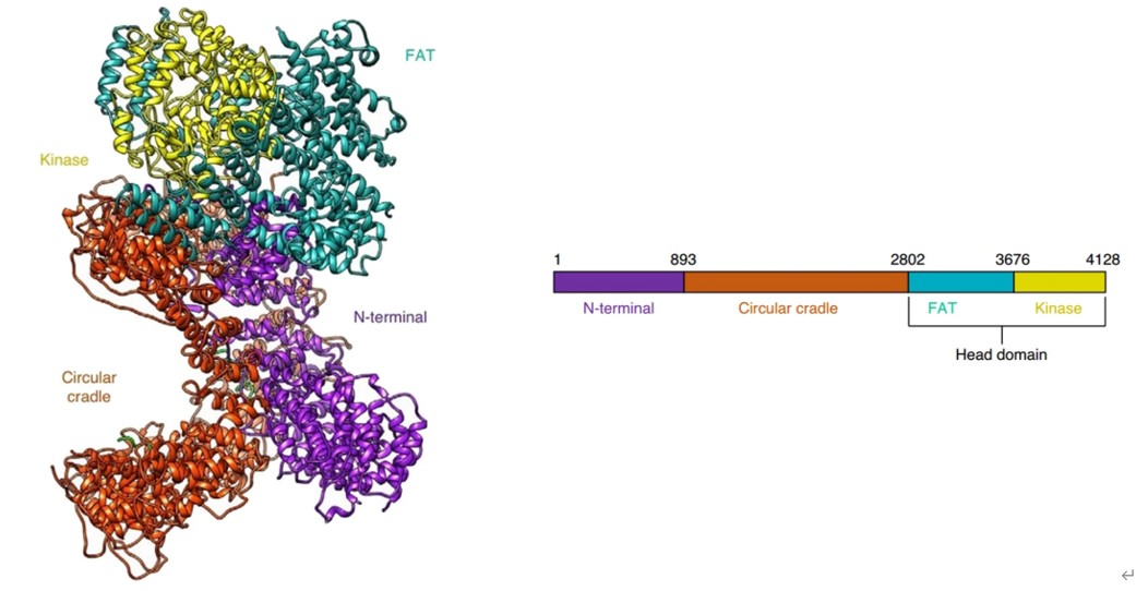 Fig. 1 The overall structure of DNA-PKcs (PRKDC).1
Key structural properties of PRKDC:
- Large ring structures containing an N-terminal HEAT repeat region and a C-terminal kinase domain
- The FAT and FATC domains form an intramolecular interaction interface to regulate kinase activity
- The kinase domain is responsible for phosphorylating downstream substrates and initiating DNA damage signaling
- The HEAT repeat region mediates protein-protein interactions with Ku70/Ku80 and other repair factors
Functions of PRKDC
The core function of PRKDC is to repair DNA double-strand breaks and maintain genomic stability. Additionally, it plays a significant role in the development of the immune system and the maintenance of telomeres.
| Function | Description |
| DNA Repair | PRKDC, as the catalytic subunit of the DNA-PK complex, repairs DNA double-strand breaks through the non-homologous end joining pathway. |
| Immune Regulation | During the V(D)J recombination process, the genes encoding immunoglobulins and T-cell receptors are cut and rearranged to generate diverse antigen receptors. |
| Telomere Maintenance | Located in the telomere region, it participates in the regulation of telomere length and the protection of the ends, preventing chromosome fusion. |
| Transcriptional Regulation | Phosphorylation of various transcription factors such as p53, and regulation of the expression of genes related to DNA damage response. |
| Cell Cycle Checkpoints | Activate cell cycle checkpoints after DNA damage to prevent damaged DNA replication and cell division. |
PRKDC, upon recognizing DNA double-strand breaks, recruits and phosphorylates downstream repair factors, initiating the damage repair cascade reaction. Its kinase activity depends on the binding to the Ku70/Ku80 complex, forming an active full enzyme complex at the DNA damage site.
Applications of PRKDC and PRKDC Antibody in Literature
1. Chen, Yu, et al. "Role of PRKDC in cancer initiation, progression, and treatment." Cancer cell international 21.1 (2021): 563. https://doi.org/10.1186/s12935-021-02229-8
The article indicates that the PRKDC gene encodes the DNA-PKcs protein, which repairs DNA double-strand breaks through non-homologous end joining and is involved in immune tolerance and chromosome stability. DNA-PKcs affects tumor occurrence and development as well as the efficacy of radiotherapy, chemotherapy and immunotherapy, and has predictive value for treatment.
2. Miao, Zhenyan, et al. "Hsa_circ_0136666 stimulates gastric cancer progression and tumor immune escape by regulating the miR-375/PRKDC Axis and PD-L1 phosphorylation." Molecular Cancer 22.1 (2023): 205. https://doi.org/10.1186/s12943-023-01883-y
The article indicates that the circular RNA hsa_circ_0136666 is highly expressed in gastric cancer. It adsorbs miR-375-3p to upregulate PRKDC, promotes the phosphorylation of PD-L1 and prevents its degradation, leading to immune escape. siRNAs targeting this axis can enhance the anti-PD-L1 efficacy.
3. Saathoff, Miranda R., et al. "LMNA-PRKDC axis enhances DNA repair and promotes chemoresistance in glioblastoma." Cell Death & Disease (2025).https://doi.org/10.1038/s41419-025-08226-3
The article indicates that the LMNA-PRKDC axis in glioblastoma enhances DNA repair, leading to temozolomide resistance. The PRKDC inhibitor KU57788 can reverse the resistance and restore chemotherapy sensitivity. This axis is highly expressed in the stem cell-like tumor subpopulation, indicating poor patient survival and making it a potential therapeutic target.
4. Chen, Yu, et al. "Prevalence of PRKDC mutations and association with response to immune checkpoint inhibitors in solid tumors." Molecular oncology 14.9 (2020): 2096-2110. https://doi.org/10.1002/1878-0261.12739
The article indicates that PRKDC gene mutations are positively correlated with high tumor mutational burden, inflammatory microenvironment, and the efficacy of immune checkpoint inhibitors. The infiltration of CD8+ T cells and other immune cells increases in the mutated samples. The survival period of patients carrying PRKDC mutations is significantly prolonged, suggesting that it can serve as a biomarker for predicting the efficacy of immunotherapy.
5. Yang, Xiawei, et al. "Potential value of PRKDC as a therapeutic target and prognostic biomarker in pan-cancer." Medicine 101.27 (2022): e29628. https://doi.org/10.1097/MD.0000000000029628
Pan-cancer analysis revealed that PRKDC is highly expressed in tumor tissues and is associated with poor prognosis in patients. Its expression level is positively correlated with tumor immune infiltration and is involved in pathways such as cell cycle. Mutations in PRKDC are associated with better survival. This gene can serve as a therapeutic target and prognostic marker for pan-cancer.
Creative Biolabs: PRKDC Antibodies for Research
Creative Biolabs specializes in the production of high-quality PRKDC antibodies for research and industrial applications. Our portfolio includes monoclonal and polyclonal antibodies tailored for ELISA, Flow Cytometry, Western blot, immunohistochemistry, and other diagnostic methodologies.
- Custom PRKDC Antibody Development: Tailor-made solutions to meet specific research requirements.
- Bulk Production: Large-scale antibody manufacturing for industry partners.
- Technical Support: Expert consultation for protocol optimization and troubleshooting.
- Aliquoting Services: Conveniently sized aliquots for long-term storage and consistent experimental outcomes.
For more details on our PRKDC antibodies, custom preparations, or technical support, contact us at email.
Reference
- Chen, Yu, et al. "Role of PRKDC in cancer initiation, progression, and treatment." Cancer cell international 21.1 (2021): 563. Distributed under Open Access license CC BY 4.0, without modification. https://doi.org/10.1186/s12935-021-02229-8
Anti-PRKDC antibodies
 Loading...


Hot products 
-
Mouse Anti-CASP7 Recombinant Antibody (10-01-62) (CBMAB-C2005-LY)

-
Mouse Anti-AK4 Recombinant Antibody (V2-180419) (CBMAB-A1891-YC)

-
Mouse Anti-ASB9 Recombinant Antibody (1D8) (CBMAB-A0529-LY)

-
Mouse Anti-COL1A2 Recombinant Antibody (CF108) (V2LY-1206-LY626)

-
Mouse Anti-ADV Recombinant Antibody (V2-503423) (CBMAB-V208-1364-FY)

-
Mouse Anti-CD33 Recombinant Antibody (6C5/2) (CBMAB-C8126-LY)

-
Mouse Anti-ATP1B1 Recombinant Antibody (E4) (CBMAB-0463-LY)

-
Mouse Anti-C5B-9 Recombinant Antibody (CBFYA-0216) (CBMAB-X0304-FY)

-
Mouse Anti-BLNK Recombinant Antibody (CBYY-0623) (CBMAB-0626-YY)

-
Mouse Anti-AAV9 Recombinant Antibody (V2-634029) (CBMAB-AP023LY)

-
Mouse Anti-4-Hydroxynonenal Recombinant Antibody (V2-502280) (CBMAB-C1055-CN)

-
Mouse Anti-DLC1 Recombinant Antibody (D1009) (CBMAB-D1009-YC)

-
Mouse Anti-EPO Recombinant Antibody (CBFYR0196) (CBMAB-R0196-FY)

-
Mouse Anti-AGK Recombinant Antibody (V2-258056) (CBMAB-M0989-FY)

-
Mouse Anti-AMACR Recombinant Antibody (CB34A) (CBMAB-CA034LY)

-
Mouse Anti-ARG1 Recombinant Antibody (CBYCL-103) (CBMAB-L0004-YC)

-
Mouse Anti-CCS Recombinant Antibody (CBFYC-1093) (CBMAB-C1150-FY)

-
Mouse Anti-ALX1 Recombinant Antibody (96k) (CBMAB-C0616-FY)

-
Rabbit Anti-ADRA1A Recombinant Antibody (V2-12532) (CBMAB-1022-CN)

-
Mouse Anti-EGR1 Recombinant Antibody (CBWJZ-100) (CBMAB-Z0289-WJ)

- AActivation
- AGAgonist
- APApoptosis
- BBlocking
- BABioassay
- BIBioimaging
- CImmunohistochemistry-Frozen Sections
- CIChromatin Immunoprecipitation
- CTCytotoxicity
- CSCostimulation
- DDepletion
- DBDot Blot
- EELISA
- ECELISA(Cap)
- EDELISA(Det)
- ESELISpot
- EMElectron Microscopy
- FFlow Cytometry
- FNFunction Assay
- GSGel Supershift
- IInhibition
- IAEnzyme Immunoassay
- ICImmunocytochemistry
- IDImmunodiffusion
- IEImmunoelectrophoresis
- IFImmunofluorescence
- IGImmunochromatography
- IHImmunohistochemistry
- IMImmunomicroscopy
- IOImmunoassay
- IPImmunoprecipitation
- ISIntracellular Staining for Flow Cytometry
- LALuminex Assay
- LFLateral Flow Immunoassay
- MMicroarray
- MCMass Cytometry/CyTOF
- MDMeDIP
- MSElectrophoretic Mobility Shift Assay
- NNeutralization
- PImmunohistologyp-Paraffin Sections
- PAPeptide Array
- PEPeptide ELISA
- PLProximity Ligation Assay
- RRadioimmunoassay
- SStimulation
- SESandwich ELISA
- SHIn situ hybridization
- TCTissue Culture
- WBWestern Blot
