FTH1 Antibodies
Background
The FTH1 gene encodes a protein called ferritin heavy chain, which is the main subunit of the ferritin complex and is mainly distributed in tissues such as the liver and spleen of vertebrates. This protein, by binding to iron ions, assumes the functions of iron storage and buffering within cells, thereby protecting cells from free iron toxicity and maintaining the balance of iron metabolism. Mammalian cells rely on FTH1 to cope with fluctuations in iron levels, as dynamic changes in iron can affect REDOX homeostasis. The structure of ferritin was first resolved in the mid-20th century. The cavity structure formed by the heavy chain and the light chain together provides a classic model for understanding bionalization and metal ion regulation. The efficient iron storage mechanism of FTH1 has been widely studied and holds significant scientific importance for exploring the functions of metalloproteins, cellular antioxidant defense, and the mechanisms of iron-related diseases.
Structure of FTH1
The molecular weight of the ferritin heavy chain subunit encoded by the FTH1 gene is approximately 21 kDa, and this molecular weight fluctuates slightly among different species due to minor differences in amino acid sequences.
| Species | Human | Mouse | Rat | Bovine |
|---|---|---|---|---|
| Molecular Weight (kDa) | 21 | 20.8 | 21 | 20.9 |
| Primary Structural Differences | With iron oxidase activity | Highly conservative functions | Similar to humans | Sequence high homology |
This protein is composed of approximately 183 amino acids and forms a typical quaternary structure by folding its primary structure. The core function of FTH1 depends on the "iron oxidase" active center it forms, which contains multiple conserved histidine and glutamic acid residues and can catalyze the oxidation of divalent iron to trivalent iron and store it in the protein cavity. Its secondary structure is mainly composed of α -helices, which entangle with each other to form a hollow spherical structure, providing a site for the deposition of iron cores. A key feature is its possession of the "Ferroxidase" active site, which is not available in the light chain subunits. It is directly responsible for the rapid oxidation of iron ions and is crucial for the iron metabolism balance and antioxidant defense of cells.
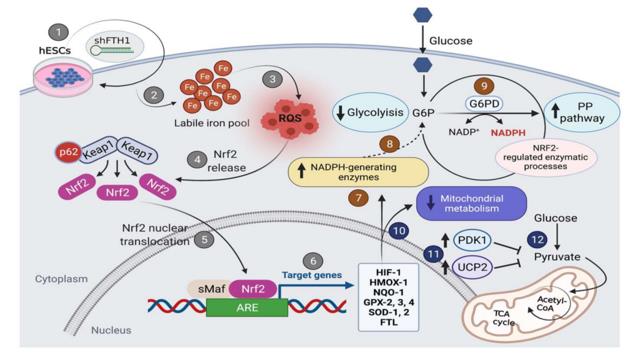 Fig. 1 Schematic diagram illustrating oxidative stress response mediated by FTH1 downregulation in hESCs.1
Key structural properties of FTH1:
- Conservative α -helical bundle tetramer structure
- Hydrophobic core constitute iron oxidase activity center
- Unique Ferroxidase loci ferrous iron oxidation
Functions of FTH1
The core function of the ferritin heavy chain encoded by the FTH1 gene is the storage and buffering of iron ions. It also participates in various cellular processes, including antioxidant defense and immune regulation.
| Function | Description |
|---|---|
| Iron storage and buffering | Bind and store excessive divalent iron ions within cells, converting them into non-toxic mineralized trivalent iron forms to prevent the Fenton reaction from generating free radicals. |
| Steady-state regulation of iron | By storing and releasing iron, it dynamically regulates the concentration of free iron pools within cells, providing an iron source for physiological processes such as hemoglobin synthesis. |
| Antioxidant protection | Effectively isolate free iron with REDOX activity, reduce the production of reactive oxygen species such as hydroxyl radicals, and protect cells from oxidative damage. |
| Immune regulation | Under inflammatory conditions, its expression is upregulated to isolate the iron required for pathogen proliferation, exerting a "nutritional immune" effect. |
| Cell proliferation support | Provide sufficient iron supply for rapidly dividing cells (such as stem cells and tumor cells) to support DNA synthesis and energy metabolism. |
Ferritin exhibits a synergistic binding to iron. Its spherical cavity structure allows for the storage of up to 4,500 iron atoms, making it an efficient and high-capacity intracellular iron buffering system, which is different from membrane proteins that directly transport iron.
Applications of FTH1 and FTH1 Antibody in Literature
1. Cui, Jingxuan, et al. "Protosappanin A protects DOX‐induced myocardial injury and cardiac dysfunction by targeting ACSL4/FTH1 axis‐dependent ferroptosis." Advanced Science 11.34 (2024): 2310227. https://doi.org/10.1002/advs.202310227
This study reports that protohematoxylin A effectively inhibits myocardial ferroptosis by targeting and binding to ACSL4 and FTH1, alleviates myocardial injury and dysfunction caused by doxorubicin, and provides a new therapeutic strategy for related cardiotoxic diseases.
2. Wang, Kun, et al. "Locally organised and activated Fth1hi neutrophils aggravate inflammation of acute lung injury in an IL-10-dependent manner." Nature Communications 13.1 (2022): 7703. https://doi.org/10.1038/s41467-022-35492-y
This study found through single-cell sequencing that in LPS-induced acute lung injury, there are two distinct neutrophil subpopulations with high expression of Fth1 and high expression of Prok2 in the lungs. Among them, the Fth1hi subgroup has stronger inflammatory and antioxidant properties. Its persistent retention will aggravate lung injury and may serve as a biomarker for the poor prognosis of ARDS.
3. Di Sanzo, Maddalena, et al. "FTH1 pseudogenes in cancer and cell metabolism." Cells 9.12 (2020): 2554. https://doi.org/10.3390/cells9122554
The article indicates that pseudogenes were once mistakenly regarded as "junk DNA", but in fact, they are defective copies of functional genes. Studies have shown that many pseudogenes play important biological roles through transcriptional regulatory mechanisms. This article focuses on reviewing the ferritin family, especially the possible functions and regulatory networks of its FTH1 pseudogene.
4. Dang, Yini, et al. "FTH1-and SAT1-induced astrocytic ferroptosis is involved in Alzheimer's disease: evidence from single-cell transcriptomic analysis." Pharmaceuticals 15.10 (2022): 1177. https://doi.org/10.3390/ph15101177
The article indicates that through single-cell sequencing analysis, it was found that astrocytes in the entorhinal cortex of patients with Alzheimer's disease decreased and the ferroptosis pathway was activated. FTH1 and SAT1 have been identified as key molecules regulating ferroptosis in astrocytes, which may be associated with emotional and cognitive impairment in AD, providing new targets for treatment.
5. Zhang, Luxia, et al. "Genome-Wide Identification and Functional Differentiation of the FTH1 Gene Family: Insights into Immune Response to Vibrio in the Blood Clam Anadara granosa." Fishes 10.12 (2025): 646. https://doi.org/10.3390/fishes10120646
The article indicates that seven FTH1 homologous genes have been identified in the genome of the clam. Among them, five contain conserved domains, whose expression is downregulated after Vibrio infection, which may be related to ferroptosi-like pathogen clearance. The remaining gene structures are atypical, and it is speculated that they are involved in non-classical functions such as vesicle transport.
Creative Biolabs: FTH1 Antibodies for Research
Creative Biolabs specializes in the production of high-quality FTH1 antibodies for research and industrial applications. Our portfolio includes monoclonal antibodies tailored for ELISA, Flow Cytometry, Western blot, immunohistochemistry, and other diagnostic methodologies.
- Custom FTH1 Antibody Development: Tailor-made solutions to meet specific research requirements.
- Bulk Production: Large-scale antibody manufacturing for industry partners.
- Technical Support: Expert consultation for protocol optimization and troubleshooting.
- Aliquoting Services: Conveniently sized aliquots for long-term storage and consistent experimental outcomes.
For more details on our FTH1 antibodies, custom preparations, or technical support, contact us at email.
Reference
- Scaramuzzino, Luana, et al. "Uncovering the metabolic and stress responses of human embryonic stem cells to FTH1 gene silencing." Cells 10.9 (2021): 2431. https://doi.org/10.3390/cells10092431
Anti-FTH1 antibodies
 Loading...


Hot products 
-
Mouse Anti-FPR2 Recombinant Antibody (1D6) (CBMAB-F2628-CQ)

-
Mouse Anti-ALOX5 Recombinant Antibody (33) (CBMAB-1890CQ)

-
Mouse Anti-CALR Recombinant Antibody (CBFYC-0763) (CBMAB-C0818-FY)

-
Mouse Anti-ATP1B1 Recombinant Antibody (E4) (CBMAB-0463-LY)

-
Mouse Anti-CD164 Recombinant Antibody (CBFYC-0077) (CBMAB-C0086-FY)

-
Mouse Anti-ATG5 Recombinant Antibody (9H197) (CBMAB-A3945-YC)

-
Mouse Anti-CCS Recombinant Antibody (CBFYC-1093) (CBMAB-C1150-FY)

-
Mouse Anti-ACLY Recombinant Antibody (V2-179314) (CBMAB-A0610-YC)

-
Mouse Anti-ADV Recombinant Antibody (V2-503423) (CBMAB-V208-1364-FY)

-
Mouse Anti-CCNH Recombinant Antibody (CBFYC-1054) (CBMAB-C1111-FY)

-
Mouse Anti-C5B-9 Recombinant Antibody (CBFYA-0216) (CBMAB-X0304-FY)

-
Mouse Anti-ASB9 Recombinant Antibody (1D8) (CBMAB-A0529-LY)

-
Mouse Anti-CD2AP Recombinant Antibody (BR083) (CBMAB-BR083LY)

-
Mouse Anti-CD59 Recombinant Antibody (CBXC-2097) (CBMAB-C4421-CQ)

-
Rabbit Anti-ABL1 (Phosphorylated Y185) Recombinant Antibody (V2-443434) (PTM-CBMAB-0001YC)

-
Mouse Anti-DLC1 Recombinant Antibody (D1009) (CBMAB-D1009-YC)

-
Mouse Anti-AKR1C3 Recombinant Antibody (V2-12560) (CBMAB-1050-CN)

-
Mouse Anti-CRTAM Recombinant Antibody (CBFYC-2235) (CBMAB-C2305-FY)

-
Mouse Anti-FLI1 Recombinant Antibody (CBXF-0733) (CBMAB-F0435-CQ)

-
Mouse Anti-CD24 Recombinant Antibody (2Q1282) (CBMAB-C1624-CN)

- AActivation
- AGAgonist
- APApoptosis
- BBlocking
- BABioassay
- BIBioimaging
- CImmunohistochemistry-Frozen Sections
- CIChromatin Immunoprecipitation
- CTCytotoxicity
- CSCostimulation
- DDepletion
- DBDot Blot
- EELISA
- ECELISA(Cap)
- EDELISA(Det)
- ESELISpot
- EMElectron Microscopy
- FFlow Cytometry
- FNFunction Assay
- GSGel Supershift
- IInhibition
- IAEnzyme Immunoassay
- ICImmunocytochemistry
- IDImmunodiffusion
- IEImmunoelectrophoresis
- IFImmunofluorescence
- IGImmunochromatography
- IHImmunohistochemistry
- IMImmunomicroscopy
- IOImmunoassay
- IPImmunoprecipitation
- ISIntracellular Staining for Flow Cytometry
- LALuminex Assay
- LFLateral Flow Immunoassay
- MMicroarray
- MCMass Cytometry/CyTOF
- MDMeDIP
- MSElectrophoretic Mobility Shift Assay
- NNeutralization
- PImmunohistologyp-Paraffin Sections
- PAPeptide Array
- PEPeptide ELISA
- PLProximity Ligation Assay
- RRadioimmunoassay
- SStimulation
- SESandwich ELISA
- SHIn situ hybridization
- TCTissue Culture
- WBWestern Blot
