IL17A Antibodies
Background
IL17A is a small molecule cytokine mainly secreted by activated T lymphocytes and is a core member of the interleukin-17 family. This protein activates downstream signaling pathways by binding to the receptor IL-17RA/RC complex, promotes the release of inflammatory mediators, and plays a key role in host defense, autoimmunity and inflammatory responses. Research has found that IL17A is significantly highly expressed in chronic inflammatory diseases such as psoriasis and rheumatoid arthritis, and its abnormal activation can lead to tissue damage. This factor was first cloned from rodent T-cell hybridoma by Rouvier's team in 1993. The analysis of its receptor mechanism has opened up a new path for the treatment of autoimmune diseases. Monoclonal antibody drugs targeting IL17A have been successfully applied in clinical practice, significantly promoting the development of immunopathology and targeted therapy fields.
Structure of IL17A
IL17A is a secretory cytokine with a molecular weight of approximately 15-20 kDa, and its mature protein is composed of 155 amino acids. This protein functions in the form of a homodimer, and its tertiary structure exhibits a typical "cysteine junction" folding pattern. The molecular characteristics of IL17A among different species are compared as follows:
| Species | Human | Mouse | Rat | Rhesus monkey |
| Molecular Weight (kDa) | 15.2 | 17.5 | 17.3 | 15.4 |
| Primary Structural Differences | Contains four conserved cysteine residues to form disulfide bonds | The degree of C-terminal glycosylation is relatively high | 85% homology with mouse | 95% homology with human sequence |
The secondary structure of IL17A is mainly composed of β -folding, forming a stable spatial conformation through intramolecular disulfide bonds. Its active center is located in the conserved region on the protein surface and activates downstream signaling pathways by specifically binding to the IL-17 receptor. The N-glycosylation modification sites of this protein vary among different species, directly affecting its biological activity and immunogenicity.
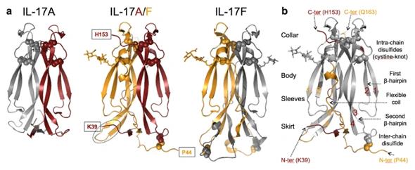 Fig. 1 Structure of human IL-17A/F.1
Key structural properties of IL17A:
- Unique "cysteine junction" folding structure
- Conformation of homologous dimer stabilized by disulfide bonds
- Conserved receptor binding interface
Functions of IL17A
The main function of IL17A is to mediate immune defense and inflammatory responses. However, this factor is also involved in various pathological processes, including the occurrence of autoimmune diseases and the regulation of tissue repair.
| Function | Description |
| Activation of immune defense | Promote the release of antimicrobial peptides and inflammatory factors by epithelial cells and fibroblasts, and enhance the host's ability to clear extracellular pathogens. |
| The inflammatory response is initiated | Induce various cells to express inflammatory mediators such as IL-6 and G-CSF, amplify local inflammatory signals, and recruit immune cells such as neutrophils. |
| Regulation of tissue barriers | Adjust epithelial tight junction protein expression, affect the mucosal barrier integrity, play a key role in the gut and the skin immunity. |
| Pathological injury mechanism | Overexpression can disrupt immune tolerance, lead to tissue inflammation and abnormal repair, and promote the progression of autoimmune diseases such as psoriasis and arthritis. |
| Participation in tissue repair | Under specific conditions, it participates in the process of wound healing and tissue remodeling by promoting the proliferation of keratinocytes and fibroblasts. |
The signal activation of IL17A is characterized by its rapid and transient nature, in sharp contrast to persistent cytokines such as interferon - γ. This reflects its functional positioning as a "initiator" of acute inflammatory responses and also explains the pathological basis that its continuous expression in chronic inflammation is prone to cause tissue damage.
Applications of IL17A and IL17A Antibody in Literature
1. Domanski, Leszek, et al. "IL17A and IL17F genes polymorphisms are associated with histopathological changes in transplanted kidney." BMC nephrology 20.1 (2019): 124. https://doi.org/10.1186/s12882-019-1308-z
This study explored the relationship between the IL17A gene polymorphism rs2275913 and the histopathological changes after kidney transplantation. Research has found that the A allele of rs2275913 is associated with more severe phlebitis, while the G allele is linked to more significant arterial hyaluronidosis and increased mesangial matrix.
2. Miller, Jessica E., et al. "IL-17A modulates peritoneal macrophage recruitment and M2 polarization in endometriosis." Frontiers in Immunology 11 (2020): 108. https://doi.org/10.3389/fimmu.2020.00108
This study explores the role of IL-17A in endometriosis. It was found that IL-17A can indirectly induce macrophages to polarize towards the M2 phenotype by acting on ectopic endometrial epithelial cells, which may promote the disease progression of endometriosis.
3. Adams, Ralph, et al. "Bimekizumab, a novel humanized IgG1 antibody that neutralizes both IL-17A and IL-17F." Frontiers in immunology 11 (2020): 1894. https://doi.org/10.3389/fimmu.2020.01894
This study developed a novel monoclonal antibody, bimekizumab, which can effectively neutralize both IL-17A and IL-17F simultaneously. Compared with secukinumab and eccilizumab, which only target IL-17A, this dual neutralization strategy shows better potential in the treatment of inflammatory diseases such as psoriasis.
4. Pawlik, Andrzej, et al. "IL17A and IL17F gene polymorphisms in patients with rheumatoid arthritis." BMC musculoskeletal disorders 17.1 (2016): 208. https://doi.org/10.1186/s12891-016-1064-1
This study explored the association between the polymorphisms of IL17A and IL17F genes and rheumatoid arthritis (RA). The results showed that in the Polish population, the IL17A (rs2275913) allele detected had no significant correlation with the susceptibility to RA or the main clinical manifestations.
5. McKelvey, Kelly J., et al. "Co-expression of CD21L and IL17A defines a subset of rheumatoid synovia, characterised by large lymphoid aggregates and high inflammation." PloS one 13.8 (2018): e0202135. https://doi.org/10.1371/journal.pone.0202135
In this study, the synovium of rheumatoid arthritis was classified into different subtypes based on the expression of IL17A and CD21L genes. Among them, the synovial subtype that is IL17A positive and accompanied by large B-cell aggregation has an inflammatory feature mainly characterized by B-cell-mediated immune responses, which provides a molecular basis for targeted and precise treatment.
Creative Biolabs: IL17A Antibodies for Research
Creative Biolabs specializes in the production of high-quality IL17A antibodies for research and industrial applications. Our portfolio includes monoclonal antibodies tailored for ELISA, Flow Cytometry, Western blot, immunohistochemistry, and other diagnostic methodologies.
- Custom IL17A Antibody Development: Tailor-made solutions to meet specific research requirements.
- Bulk Production: Large-scale antibody manufacturing for industry partners.
- Technical Support: Expert consultation for protocol optimization and troubleshooting.
- Aliquoting Services: Conveniently sized aliquots for long-term storage and consistent experimental outcomes.
For more details on our IL17A antibodies, custom preparations, or technical support, contact us at email.
Reference
- Goepfert, Arnaud, et al. "The human IL-17A/F heterodimer: a two-faced cytokine with unique receptor recognition properties." Scientific reports 7.1 (2017): 8906. https://doi.org/10.1038/s41598-017-08360-9
Anti-IL17A antibodies
 Loading...


Hot products 
-
Human Anti-SARS-CoV-2 Spike Recombinant Antibody (CR3022) (CBMAB-CR014LY)

-
Mouse Anti-ALX1 Recombinant Antibody (96k) (CBMAB-C0616-FY)

-
Mouse Anti-BMI1 Recombinant Antibody (CBYC-P026) (CBMAB-P0108-YC)

-
Mouse Anti-ACKR3 Recombinant Antibody (V2-261265) (CBMAB-C1023-LY)

-
Mouse Anti-CD8 Recombinant Antibody (C1083) (CBMAB-C1083-LY)

-
Mouse Anti-AAV8 Recombinant Antibody (V2-634028) (CBMAB-AP022LY)

-
Mouse Anti-ANXA7 Recombinant Antibody (A-1) (CBMAB-A2941-YC)

-
Mouse Anti-HTLV-1 gp46 Recombinant Antibody (CBMW-H1006) (CBMAB-V208-1154-FY)

-
Rat Anti-ABCC11 Recombinant Antibody (V2-179001) (CBMAB-A0236-YC)

-
Mouse Anti-AKT1/AKT2/AKT3 (Phosphorylated T308, T309, T305) Recombinant Antibody (V2-443454) (PTM-CBMAB-0030YC)

-
Mouse Anti-DHFR Recombinant Antibody (D0821) (CBMAB-D0821-YC)

-
Mouse Anti-8-oxoguanine Recombinant Antibody (V2-7697) (CBMAB-1869CQ)

-
Mouse Anti-Acetyl SMC3 (K105/K106) Recombinant Antibody (V2-634053) (CBMAB-AP052LY)

-
Mouse Anti-CFL1 Recombinant Antibody (CBFYC-1771) (CBMAB-C1833-FY)

-
Mouse Anti-C5b-9 Recombinant Antibody (aE11) (CBMAB-AO138LY)

-
Mouse Anti-ABIN2 Recombinant Antibody (V2-179106) (CBMAB-A0349-YC)

-
Mouse Anti-CECR2 Recombinant Antibody (CBWJC-2465) (CBMAB-C3533WJ)

-
Mouse Anti-A2M Recombinant Antibody (V2-178822) (CBMAB-A0036-YC)

-
Mouse Anti-CCND2 Recombinant Antibody (DCS-3) (CBMAB-G1318-LY)

-
Mouse Anti-ARSA Recombinant Antibody (CBYC-A799) (CBMAB-A3679-YC)

- AActivation
- AGAgonist
- APApoptosis
- BBlocking
- BABioassay
- BIBioimaging
- CImmunohistochemistry-Frozen Sections
- CIChromatin Immunoprecipitation
- CTCytotoxicity
- CSCostimulation
- DDepletion
- DBDot Blot
- EELISA
- ECELISA(Cap)
- EDELISA(Det)
- ESELISpot
- EMElectron Microscopy
- FFlow Cytometry
- FNFunction Assay
- GSGel Supershift
- IInhibition
- IAEnzyme Immunoassay
- ICImmunocytochemistry
- IDImmunodiffusion
- IEImmunoelectrophoresis
- IFImmunofluorescence
- IGImmunochromatography
- IHImmunohistochemistry
- IMImmunomicroscopy
- IOImmunoassay
- IPImmunoprecipitation
- ISIntracellular Staining for Flow Cytometry
- LALuminex Assay
- LFLateral Flow Immunoassay
- MMicroarray
- MCMass Cytometry/CyTOF
- MDMeDIP
- MSElectrophoretic Mobility Shift Assay
- NNeutralization
- PImmunohistologyp-Paraffin Sections
- PAPeptide Array
- PEPeptide ELISA
- PLProximity Ligation Assay
- RRadioimmunoassay
- SStimulation
- SESandwich ELISA
- SHIn situ hybridization
- TCTissue Culture
- WBWestern Blot
